Ankle
The ankle, the talocrural region[1] or the jumping bone (informal) is the area where the foot and the leg meet.
In medical terminology, "ankle" (without qualifiers) can refer broadly to the region or specifically to the talocrural joint.
[1][6] The main bones of the ankle region are the talus (in the foot), the tibia, and fibula (both in the leg).
[11] The superior extensor retinaculum of foot extends between the anterior (forward) surfaces of the tibia and fibula near their lower (distal) ends.
The flexor retinaculum of foot extends from the medial malleolus to the medical process of the calcaneus, and the following structures in order from medial to lateral: the tendon of the tibialis posterior muscle, the tendon of the flexor digitorum longus muscle, the posterior tibial artery and vein, the tibial nerve, and the tendon of the flexor hallucis longus muscle.
The superior fibular retinaculum extends from the deep transverse fascia of the leg and lateral malleolus to calcaneus.
[9]: 1418–9 Mechanoreceptors of the ankle send proprioceptive sensory input to the central nervous system (CNS).
[15] Muscle spindles are thought to be the main type of mechanoreceptor responsible for proprioceptive attributes from the ankle.
This was done by using a fMRI machine in order to see the changes in brain activity when the receptors of the ankle are stimulated.
Historically, the role of the ankle in locomotion has been discussed by Aristotle and Leonardo da Vinci.
There is no question that ankle push-off is a significant force in human gait, but how much energy is used in leg swing as opposed to advancing the whole-body center of mass is not clear.
[20] Complications may include an associated high ankle sprain, compartment syndrome, stiffness, malunion, and post-traumatic arthritis.
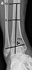
Varus or valgus deformity, if suspected, can be measured with the frontal tibiotalar surface angle (TTS), formed by the mid-longitudinal tibial axis (such as through a line bisecting the tibia at 8 and 13 cm above the tibial plafond) and the talar surface.
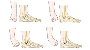
Loss of any of these normal anatomic spaces can indirectly reflect ligamentous injury or occult fracture, and can be followed by MRI or CT.[26] Clubfoot or talipes equinovarus, which occurs in one to two of every 1,000 live births, involves multiple abnormalities of the foot.
[27] Equinus refers to the downard deflection of the ankle, and is named for the walking on the toes in the manner of a horse.
[28] This does not occur because it is accompanied by an inward rotation of the foot (varus deformity), which untreated, results in walking on the sides of the feet.
[31] The word ankle or ancle is common, in various forms, to Germanic languages, probably connected in origin with the Latin angulus, or Greek αγκυλος, meaning bent.