Autonomic nervous system
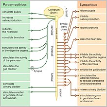
An older simplification of the sympathetic and parasympathetic nervous systems as "excitatory" and "inhibitory" was overturned due to the many exceptions found.
A third subsystem of neurons has been named as non-noradrenergic, non-cholinergic transmitters (because they use nitric oxide as a neurotransmitter) and are integral in autonomic function, in particular in the gut and the lungs.
[10] Although the ANS is also known as the visceral nervous system and although most of its fibers carry non-somatic information to the CNS, many authors still consider it only connected with the motor side.
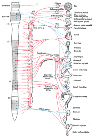
The parasympathetic nervous system consists of cells with bodies in one of two locations: the brainstem (cranial nerves III, VII, IX, X) or the sacral spinal cord (S2, S3, S4).
Concurrently, the sacral section of the neural crest provides an additional layer of complexity by contributing input to the hindgut ganglia.
Throughout this developmental journey, numerous receptors exhibiting tyrosine kinase activity, such as Ret and Kit, play indispensable roles.
Ret, for instance, plays a critical role in the formation of enteric ganglia derived from cells known as vagal neural crest.
This description is rooted in the ENS's ability to communicate independently with the central nervous system through parasympathetic and sympathetic neurons.
Additionally, the myenteric plexus plays a unique role in innervating motor end plates with the inhibitory neurotransmitter nitric oxide in the striated-muscle segment of the esophagus, a feature exclusive to this organ.
Resembling the astrocytes of the central nervous system, enteric glial cells respond to cytokines by expressing MHC class II antigens and generating interleukins.
This underlines their pivotal role in modulating inflammatory responses in the intestine, adding another layer of sophistication to the functional dynamics of the ENS.
The rich structural diversity of enteric neurons highlights the complexity and adaptability of the ENS in orchestrating a wide array of gastrointestinal functions, reflecting its status as a dynamic and sophisticated component of the nervous system.
These sensory neurons monitor the levels of carbon dioxide, oxygen and sugar in the blood, arterial pressure and the chemical composition of the stomach and gut content.
The nTS also receives input from a nearby chemosensory center, the area postrema, that detects toxins in the blood and the cerebrospinal fluid and is essential for chemically induced vomiting or conditional taste aversion (the memory that ensures that an animal that has been poisoned by a food never touches it again).
For example, standing up from a reclining or sitting position would entail an unsustainable drop in blood pressure if not for a compensatory increase in the arterial sympathetic tonus.
Another example is the constant, second-to-second, modulation of heart rate by sympathetic and parasympathetic influences, as a function of the respiratory cycles.
Higher organisms maintain their integrity via homeostasis which relies on negative feedback regulation which, in turn, typically depends on the autonomic nervous system.
[18] Promotes a fight-or-flight response, corresponds with arousal and energy generation, and inhibits digestion The pattern of innervation of the sweat gland—namely, the postganglionic sympathetic nerve fibers—allows clinicians and researchers to use sudomotor function testing to assess dysfunction of the autonomic nervous systems, through electrochemical skin conductance.
Recent studies indicate that ANS activation is critical for regulating the local and systemic immune-inflammatory responses and may influence acute stroke outcomes.
[citation needed] In 1665, Thomas Willis used the terminology, and in 1900, John Newport Langley used the term, defining the two divisions as the sympathetic and parasympathetic nervous systems.
In one study, caffeine provoked a greater maximum heart rate while a strenuous task was being performed compared to a placebo.
It is important to note that the data supporting increased parasympathetic activity in the supine position was derived from an experiment involving participants between the ages of 25 and 30 who were considered healthy and sedentary.