Berghia stephanieae
[4] This is one of the most commonly sold aeolid nudibranchs in the marine aquarium trade in North America,[5] because it is used to control the sea anemone Aiptasia.
The larval nervous system of Berghia stephanieae includes an apical organ, developing central ganglia, and peripheral neurons associated with the velum (a structure used for swimming and particulate food collection), foot and posterior, visceral part of the larva.
[6] The same developmental pattern of cephalic tentacles has been shown in three other nudibranchs, so far (Adalaria proxima, Cadlina laevis and Melibe leonina).
[6] The first detectable structures in the early veliger stage (5-10% of development), the larval shell and the ciliated velar lobes, appear at the same time as the first movements of the larvae (rotation around their anterior-posterior axes).
[6] Veliger stage (10-20% of development): The embryo can retract the velum into the shell and the eyes as well as the larval foot (propodium) appear.
[6] Late veliger stage (20-25% of development): The operculum is present and the foot becomes thicker and longer, the embryo hatches shortly prior to metamorphosis.
Generally, 48 hours after metamorphosis juvenile specimens of Berghia stephanieae start to prey upon pieces of Aiptasia pallida anemones.
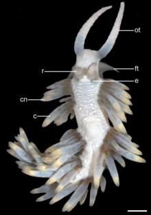
As the development continues, the length and the thickness of the rhinophores and oral tentacles increases as well as the body size.
The larval nervous system of Berghia stephanieae includes an apical organ, developing central ganglia, and peripheral neurons associated with the velum, foot and posterior part of the larvae.
Because Berghia stephanieae only eat Aiptasias, the nudibranchs will die of starvation when all the anemones are gone, so this situation must be taken into account.
This rapid and dramatic response involves the coordinated movement of cerata, which are venomous appendages protruding from the animal's mantle.
This suggests that the bristling behavior is stereotyped but non-reflexive, with the cerebral ganglia playing a crucial role in coordinating the full defensive response.


zy = zygotes , * (asterisk) = four-celled embryos within the same egg mass, cap = the capsule that surrounds each embryo.

s = shell ,
v = ciliated velum,
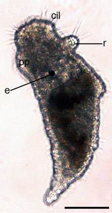
cil = cilia ,
cap = the capsule that surrounds each embryo.

| - (purple) v = cilia of apical tuft and velum | |
| - ( cornflower blue ) the larval retractor muscle, | |
| - (yellow) the accessory retractor muscle, | |
| - (red) the velar ring muscles, |
cap = the capsule that surrounds each embryo.
The total size is about 170 μm.

| - (purple) v = cilia of apical tuft and velum | |
| - (cornflower blue) the larval retractor muscle, | |
| - (yellow) the accessory retractor muscle, | |
| - (red) the velar ring muscles, | |
| - (dark blue) pedal retractor muscle, | |
| - (green) metapodial retractor muscle, |
s = shell.
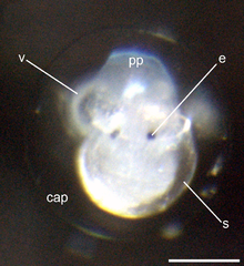
e = eyes,
pp = elaborated larval foot (propodium),
s = shell,
v = ciliated velum,
cap = the capsule that surrounds each embryo.

o = operculum ,
mp = metapodium to where is the operculum attached at,
cil = cilia,
s = shell,
v = ciliated velum.

- (open triangles) degenerating velar lobes and muscles,

| - (purple) v = cilia of apical tuft and velum | |
| - (cornflower blue) the larval retractor muscle, | |
| - (red) the velar ring muscles, | |
| - (dark blue) pedal retractor muscle, | |
| - (green) metapodial retractor muscle, |
o = operculum,
s = shell.
The total size is about 120 μm.

e = eyes,
cil = cilia,
s = shell.

j = juvenile of Berghia stephanieae is crawling out of the shell and marking the end of the metamorphosis.
pp = elaborated larval foot (propodium),
e = eyes,
s = shell.

| - (light green) longitudinal muscle fibers, | |
| - (black) circular muscle fibers, | |
| - (white) oblique muscle fibres, | |
| - (brown) anlage of the buccal musculature, | |
| - ( apricot color ) mouth, | |
| - (cornflower blue) the larval retractor muscle, |
e = eye.
The total size is about 200 μm.
r = anlage of the rhinophores,
cil = cilia. The body is covered by cilia.
e = eye,
pp = propodium.

| - (light green) longitudinal muscle fibers, | |
| - (black) circular muscle fibers, | |
| - (white) oblique muscle fibres, | |
| - (brown) anlage of the buccal musculature, | |
| - (apricot color) mouth, |
r = rhinophores,
c = the first cerata pairs,
e = eye.
The total size is about 600 μm.

ot = oral tentacles,
r = rhinophores,
c = anlagen of the first cerata .

cn =cnidosacs at the cerata tips.
Note that the rhinophores as well as oral tentacles are longer and thicker now.
ot = oral tentacles,
r = rhinophores,
e = eye,
c = cerata.

ft = foot tentacles
A tentacle-like elongation of the propodium, appear. Note that the oral tentacles are almost twice as long as the rhinophores and the additional pairs of cerata.
ot = oral tentacles,
r = rhinophores,
e = eye,
c = cerata.
cn =cnidosacs at the cerata tips.

| - (light green) longitudinal muscle fibers, | |
| - (black) circular muscle fibers, | |
| - (white) oblique muscle fibres, | |
| - (brown) anlage of the buccal musculature, | |
| - (apricot color) mouth, |
ft = foot tentacle, a tentacle-like elongation of the propodium,
ot = the anlage of oral tentacles,
r = rhinophores,
c = the first cerata pairs,
e = eye.
ot = oral tentacles,
ft = foot tentacles,
e = eye,
r = rhinophores,
cn =cnidosacs at the cerata tips,
c = cerata.