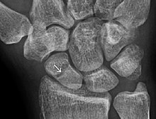
Bone healing
The healing process is mainly determined by the periosteum (the connective tissue membrane covering the bone).
The periosteum is one source of precursor cells that develop into chondroblasts and osteoblasts that are essential to the healing of bone.
Other sources of precursor cells are the bone marrow (when present), endosteum, small blood vessels, and fibroblasts.
Such healing requires only the remodeling of lamellar bone, the Haversian canals and the blood vessels without callus formation.
In this case, cutting cones, which consists of osteoclasts, form across the fracture lines, generating cavities at a rate of 50–100 μm/day.
Within a few hours, the extravascular blood cells form a clot called a hematoma[5] that acts as a template for callus formation.
These cells, including macrophages, release inflammatory mediators such as cytokines (tumor necrosis factor alpha (TNFα), interleukin-1 family (IL-1), interleukin 6 (IL-6), 11 (IL-11), and 18 (IL-18)) and increase blood capillary permeability.
Within 7–14 days, they form a loose aggregate of cells, interspersed with small blood vessels, known as granulation tissue.
[citation needed] Osteoclasts move in to reabsorb dead bone ends, and other necrotic tissue is removed.
The periosteal cells proximal to (on the near side of) the fracture gap develop into chondroblasts, which form hyaline cartilage.
[citation needed] Remodeling begins as early as three to four weeks after fracture and may take 3 to 5 years to complete.
The trabecular bone is first resorbed by osteoclasts, creating a shallow resorption pit known as a "Howship's lacuna".
[4] This process can be enhanced by certain synthetic injectable biomaterials, such as Cerament, which are osteoconductive and promote bone healing.


