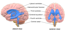
Ventricular system
The ventricular system is continuous with the central canal of the spinal cord from the fourth ventricle,[3] allowing for the flow of CSF to circulate.
[3][4] All of the ventricular system and the central canal of the spinal cord are lined with ependyma, a specialised form of epithelium connected by tight junctions that make up the blood–cerebrospinal fluid barrier.
The fourth ventricle narrows at the obex (in the caudal medulla), to become the central canal of the spinal cord.
The three primary brain vesicles represent different components of the central nervous system: the prosencephalon, mesencephalon and rhombencephalon.
[9] Separating the anterior horns of the lateral ventricles is the septum pellucidum: a thin, triangular, vertical membrane which runs as a sheet from the corpus callosum down to the fornix.
[10] The ventricles are filled with cerebrospinal fluid (CSF) which bathes and cushions the brain and spinal cord within their bony confines.
CSF is produced by modified ependymal cells of the choroid plexus found in all components of the ventricular system except for the cerebral aqueduct and the posterior and anterior horns of the lateral ventricles.
This allows the brain to grow in size and weight without resting on the floor of the cranium, which would destroy nervous tissue.
Researchers found that individuals with schizophrenia had (in terms of group averages) larger than usual ventricles.
This became the first "evidence" that schizophrenia was biological in origin and led to a renewed interest in its study via the use of imaging techniques.
Magnetic resonance imaging (MRI) has superseded the use of CT in research in the role of detecting ventricular abnormalities in psychiatric illness.
Enlarged ventricles are also found in organic dementia and have been explained largely in terms of environmental factors.