Sternum
: sternums or sterna) or breastbone is a long flat bone located in the central part of the chest.
The top of the sternum supports the clavicles (collarbones) and its edges join with the costal cartilages of the first two pairs of ribs.
Between the depression for the first costal cartilage and the demi-facet for the second is a narrow, curved edge, which slopes from above downward towards the middle.
It is flat on the front, directed upward and forward, and marked by three transverse ridges which cross the bone opposite the third, fourth, and fifth articular depressions.
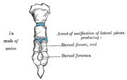
At the junction of the third and fourth parts of the body is occasionally seen an orifice, the sternal foramen, of varying size and form.
The posterior surface, slightly concave, is also marked by three transverse lines, less distinct, however, than those in front; from its lower part, on either side, the transversus thoracis takes origin.
The inferior angle has a small facet, which, with a corresponding one on the xiphoid process, forms a notch for the cartilage of the seventh rib.
These articular depressions are separated by a series of curved interarticular intervals, which diminish in length from above downward, and correspond to the intercostal spaces.
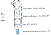
Most of the cartilages belonging to the true ribs, articulate with the sternum at the lines of junction of its primitive component segments.
[1] The sternum is composed of highly vascular tissue, covered by a thin layer of compact bone which is thickest in the manubrium between the articular facets for the clavicles.
The costal cartilage of the second rib articulates with the sternum at the sternal angle making it easy to locate.
[3] The transversus thoracis muscle is innervated by one of the intercostal nerves and superiorly attaches at the posterior surface of the lower sternum.
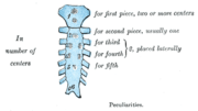
[5] These two bars fuse together along the middle to form the cartilaginous sternum which is ossified from six centers: one for the manubrium, four for the body, and one for the xiphoid process.
[citation needed] Occasionally some of the segments are formed from more than one center, the number and position of which vary [Fig.
7], or of the vertical fissure which occasionally intersects this part of the bone constituting the malformation known as fissura sterni; these conditions are further explained by the manner in which the cartilaginous sternum is formed.
When this takes place, however, the bony tissue is generally only superficial, the central portion of the intervening cartilage remaining unossified.
In particular, patients with a high BMI (obese or grossly overweight) may present with excess tissue that makes access to traditional marrow biopsy sites such as the pelvis difficult.
[14] The breastbone is sometimes cut open (a median sternotomy) to gain access to the thoracic contents when performing cardiothoracic surgery.
Such complications often entail issues like dehiscence and sternal non-union, primarily stemming from lateral forces exerted during post-operative activities such as coughing and sneezing.
The sternum can be totally removed (resected) as part of a radical surgery, usually to surgically treat a malignancy, either with or without a mediastinal lymphadenectomy (Current Procedural Terminology codes # 21632 and # 21630, respectively).
[15] The sternum, in vertebrate anatomy, is a flat bone that lies in the middle front part of the rib cage.
In birds, it is a relatively large bone and typically bears an enormous projecting keel to which the flight muscles are attached.