Nasal cartilages
These associations create vent-like structures within the nose so that air can flow from the nasal cavity to the lungs or vice versa.
Therefore, the nasal cartilages are structures that aid the body in respiratory functions to intake oxygen or expire carbon dioxide.
Surgical techniques have been produced to adjust the position or repair the nasal cartilages so that maximal airflow is once again accomplished.
[3] The septal nasal cartilage fits in a place between the perpendicular plate of the ethmoid and vomer bones while also being covered by an internal mucous membrane.
Providing two cavities generates turbulence within the tight spaces, allowing air to flow quicker bidirectionally.
[4] This can lead to respiratory issues due to low oxygen but high carbon dioxide counts within the body.
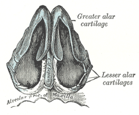
Both sides of the major alar cartilages merge to form a notch at the tip, which is referred to as the apex of the nose.
With the formation of the medial and lateral walls within the nares, the major alar cartilages function to hold open each naris.
Due to weakness corresponding with the lateral crus in certain individuals, a technique called sliding alar cartilage (SAC) has been a procedure practiced to restructure and support the nasal tip.
This associated organ plays an important role in the sense of smell by being lined with similar epithelium to that of the olfactory region of the nose.
With emerging technological advancements, reconstruction and surgical techniques have been developed to adjust the lifestyle and health of individuals.
[11] Surgery is also permitted to individuals that seek cosmetic changes due to moderate cases of a deviated septum.
Surgery may require a surgeon to cut and remove parts of the septal nasal cartilages, replacing them later in a reconstructed format.
These risks include a change in the shape of the nose, excessive bleeding, vacant space in the septum, trouble smelling, blood clots that need to be removed, and numbness by the facial region.
This open rhinoplastic procedure allows the nose to heal to an optimal position without the permanent use of man-made hardware.
In that timeframe, the tip of the nose is cut open, the greater alar cartilage is manipulated to preserve the scroll area, providing strength and structure, then the incision is sutured back up.