Cladoselache
As an early chondrichthyan, it had yet to evolve traits of modern sharks such as accelerated tooth replacement, a loose jaw suspension, enameloid teeth, and possibly claspers.
Some 20th century studies considered Cladoselache to be a basal (early-diverging) member of Elasmobranchii, the fork of cartilaginous fish which leads to modern sharks and rays.
[2][3][4] These newly identified features support a close relationship to symmoriiforms, a small group of bizarre chondrichthyans such as the bristle-spined Stethacanthus.
Cladoselache and symmoriiforms may be more closely related to chimaeras (a modern group of unusual deep-sea fish) than to true sharks and rays.
[5] Growing to several meters in length, Cladoselache is considered to have been a fast-moving and fairly agile marine predator due to its streamlined body and deeply forked tail.
From both an anatomical and historical perspective, is one of the best known of the early chondrichthyans in part due to an abundance of well-preserved fossils, discovered in the Cleveland Shale on the south shore of Lake Erie.
In addition to the cartilaginous skeleton, the fossils were so well preserved that they included traces of skin, muscle fibers, and internal organs, such as the kidneys.
This excavation contains a pair of closely spaced openings, where the two lateral branches of the dorsal aorta (a major artery) enter the braincase.
[12] Amphistyly is common in early chondrichthyans, but it is only present in hexanchiforms (frilled and sixgill sharks) among modern members of the group.
Many traits of Cladoselache’s pectoral girdle are characteristic of symmoriiforms, including large paired procoracoids, proximal radials, and a distinct metapterygium at the rear of the coracoid.
Cladoselache also has a unique trait when compared to other Paleozoic chondrichthyans: the rear edge of the metapterygium does not send out a string of small cartilage structures known as axials.
This may be due to its reduced pelvic region, a reliance on external fertilization,[11] or simply a lack of male fossils preserving the area.
[13] Cladoselache is considered one of the best-known early members of the Chondrichthyes: cartilaginous fish such as sharks, rays, and the deep-sea chimaeras (also known as ratfish or ghostsharks).
[21][1] Though it resembles modern sharks (selachians) in ecology and body shape, it is not a member of that group, which did not evolve until the Jurassic Period.
When first described near the end of the 19th century, Cladoselache was immediately recognized as a plesiomorphic (ancestral) form of cartilaginous fish, and not closely related to modern sharks despite its similar appearance.
It and other Paleozoic “sharks” may instead be described as basal chondrichthyans, a term indicating their origin at an earlier stage in cartilaginous fish evolution.
The other branch, Elasmobranchii, is much more diverse in the present, including modern sharks (Selachii), rays (Batoidea), and their extinct relatives.
[21][1] The earliest publications regarding the genus noted that both Cladoselache and acanthodians (“spiny sharks”) were close to the base of “Elasmobranchii” (Chondrichthyes), but also that both taxa carried unusual specializations relative to the estimated ancestral condition.
Cladoselache and Acanthodii were allied under the now-antiquated group Pleuropterygii, according to proposed plesiomorphic traits such as scleral rings, flap-like paired fins, a reinforced upper tail lobe, and the supposed lack of claspers.
[11] Pleuropterygii was later expanded to incorporate ctenacanthids and symmoriids, which were similar to Cladoselache in geological age, cladodont tooth shape, and general anatomy.
[13] A few studies even suggested that it was a more derived elasmobranch close to hybodonts and neoselachians (modern sharks and rays),[21][23] though this was an uncertain and unstable position.
[24][25][26][5] The dorsal spine of Cladoselache appears to be homologous to the unusual head structures prevalent in symmoriiforms,[3] and many similarities are also present in the braincase and pectoral region.
[24][25][26] However, an alternative hypothesis has caught traction: Symmoriiforms (including Cladoselache) may lie on the opposite side of Chondrichthyes, as an early part of the holocephalan stem-group which would eventually produce modern chimaeras.
[25][5] This hypothesis was originally supported by certain tentative features (reduced scalation, absent second dorsal fin spine),[25] and later stabilized further by braincase-related traits such as an arched midbrain and large orbits.
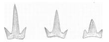
Major groups are bolded:[29] Gladbachus Gydoselache Pucapampella Doliodus Cladoselache Ozarcus Dwykaselachus Ferromirum Gutturensis Cobelodus Akmonistion Damocles Iniopera Kawichthys Helodus Debeerius Chondrenchelys Chimaeroidei (chimaeras) Elasmobranchii (true sharks, rays, and kin) The teeth of Cladoselache were slender and smooth-edged, making them suitable for grasping, but not tearing or chewing.
[17] Members of the genus were predators, and the well-preserved fossils found in the Cleveland Shale revealed a significant amount regarding their eating habits.
[35] The eye was protected by a ring of numerous (more than 20) small dermal ossifications, which were initially interpreted as denticles with a function similar to modern scleral ossicles.
This suggests that the body cavity of Cladoselache (including the digestive and urinary systems) extends further back into the caudal peduncle relative to modern sharks.
Gut content and coprolites are strongly twisted, arguing that the digestive system of Cladoselache was similar to living sharks.