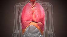
Thoracic diaphragm
Other mammals have diaphragms, and other vertebrates such as amphibians and reptiles have diaphragm-like structures, but important details of the anatomy may vary, such as the position of the lungs in the thoracic cavity.
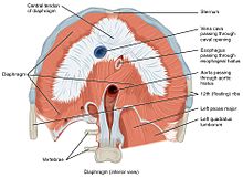
The diaphragm is an upward curved, c-shaped structure of muscle and fibrous tissue that separates the thoracic cavity from the abdomen.
[7] Its peripheral part consists of muscular fibers that take origin from the circumference of the inferior thoracic aperture and converge to be inserted into a central tendon.
The median arcuate ligament arises from the fibrous parts of right and left crura where descending thoracic aorta passes behind it.
[8] The central tendon of the diaphragm is a thin but strong aponeurosis near the center of the vault formed by the muscle, closer to the front than to the back of the thorax.
The septum transversum, the primitive central tendon of the diaphragm, originates at the rostral pole of the embryo and is relocated during longitudinal folding to the ventral thoracic region.
The pleuroperitoneal membrane and body wall myoblasts, from somatic lateral plate mesoderm, meet the septum transversum to close off the pericardio-peritoneal canals on either side of the presumptive esophagus, forming a barrier that separates the peritoneal and pleuropericardial cavities.
As the septum transversum descends inferiorly, the phrenic nerve follows, accounting for its circuitous route from the upper cervical vertebrae, around the pericardium, finally to innervate the diaphragm.
In other words, the diaphragm's movement downwards creates a partial vacuum in the thoracic cavity, which forces the lungs to expand to fill the void, drawing air in the process.
When the diaphragm relaxes (moves in the superior direction), air is exhaled by elastic recoil process of the lung and the tissues lining the thoracic cavity.
Diaphragm dysfunction is a well-known factor associated with various complications in patients, such as prolonged respiratory failure, difficulties in weaning from mechanical ventilation, extended hospitalization, increased morbidity, and mortality.
[15] Studies have reported that a thin diaphragm leads to greater lung compliance, which can contribute to respiratory failure.
In some non-human animals, the diaphragm is not crucial for breathing; a cow, for instance, can survive fairly asymptomatically with diaphragmatic paralysis as long as no massive aerobic metabolic demands are made of it.
[citation needed] If either the phrenic nerve, cervical spine or brainstem is damaged, this will sever the nervous supply to the diaphragm.
The contents of the abdomen, including the intestines, may be present in the thorax, which may impact development of the growing lungs and lead to hypoplasia.
[20] The adoption of a deeper breathing pattern typically occurs during physical exercise in order to facilitate greater oxygen absorption.
This is especially evident during deep breathing where its generally lower position increases intra-abdominal pressure, which serves to strengthen the lumbar spine.
[23][better source needed] The key to real core stabilization is to maintain the increased IAP while going through normal breathing cycles.
[23][better source needed] Therefore, if a person's diaphragm position is lower in general, through deep breathing, then this assists the strengthening of their core during that period.
Thus the model organism, the marine chordate lancelet, possesses an atriopore by which water exits the pharynx, which has been claimed (and disputed) to be homologous to structures in ascidians and hagfishes.
[25] The tunicate epicardium separates digestive organs from the pharynx and heart, but the anus returns to the upper compartment to discharge wastes through an outgoing siphon.
Thus the diaphragm emerges in the context of a body plan that separated an upper feeding compartment from a lower digestive tract, but the point at which it originates is a matter of definition.
They rely on a rocking motion of the keel of the sternum to create local areas of reduced pressure to supply thin, membranous airsacs cranially and caudally to the fixed-volume, non-expansive lungs.