Herpes simplex keratitis
[2] Primary infection typically presents as swelling of the conjunctiva and eyelids (blepharoconjunctivitis), accompanied by small white itchy lesions on the corneal surface.
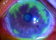
The effect of the lesions varies, from minor damage to the epithelium (superficial punctate keratitis), to more serious consequences such as the formation of dendritic ulcers.
Additional symptoms include dull pain deep inside the eye, mild to acute dryness, and sinusitis.
Subsequent recurrences may be more severe, with infected epithelial cells showing larger dendritic ulceration, and lesions forming white plaques.
[3] The epithelial layer is sloughed off as the dendritic ulcer grows, and mild inflammation (iritis) may occur in the underlying stroma of iris.
Sensation loss occurs in lesional areas, producing generalised corneal anaesthesia with repeated recurrences.
[4] The global incidence (rate of new disease) of herpes keratitis is roughly 1.5 million, including 40,000 new cases of severe monocular visual impairment or blindness each year.
Laboratory tests are indicated in complicated cases when the clinical diagnosis is uncertain and in all cases of suspected neonatal herpes infection:[4][6] Treatment of herpes of the eye is different based on its presentation: epithelial keratitis is caused by live virus while stromal disease is an immune response and metaherpetic ulcer results from inability of the corneal epithelium to heal.
[9] Oral acyclovir is as effective as topical antivirals for treating epithelial keratitis, and it has the advantage of no eye surface toxicity.
[9] Valacyclovir, a pro-drug of acyclovir likely to be just as effective for ocular disease, can cause thrombotic thrombocytopenic purpura/Hemolytic-uremic syndrome in severely immunocompromised patients such as those with AIDS; thus, it must be used with caution if the immune status is unknown.
[6] Treatment includes artificial tears and eye lubricants, stopping toxic medications, performing punctal occlusion, bandage contact lens and amniotic membrane transplant.