Histopathology
Histopathology (compound of three Greek words: ἱστός histos 'tissue', πάθος pathos 'suffering', and -λογία -logia 'study of') is the microscopic examination of tissue in order to study the manifestations of disease.
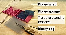
Certain specimens (especially biopsies) can undergo agar pre-embedding to assure correct tissue orientation in cassette & then in the block & then on the diagnostic microscopy slide.
[1] This process is needed to provide a properly oriented sample sturdy enough for obtaining a thin microtome section(s) for the slide.
This is usually done by hand and is a skilled job (histotechnologist) with the lab personnel making choices about which parts of the specimen microtome wax ribbon to place on slides.
In this method, the tissue is frozen and sliced thinly using a microtome mounted in a below-freezing refrigeration device called the cryostat.
A commonly performed histochemical technique is the Perls' Prussian blue reaction, used to demonstrate iron deposits in diseases like Hemochromatosis.
Called immunohistochemistry, this technique has greatly increased the ability to specifically identify categories of cells under a microscope.
The histological slides are examined under a microscope by a pathologist, a medically qualified specialist who has completed a recognised training program.
This medical diagnosis is formulated as a pathology report describing the histological findings and the opinion of the pathologist.
Scanning of slides allows for various methods of digital pathology, including the application of artificial intelligence for interpretation.
After 12 hours, there can be seen karyopyknosis and hypereosinophilia of myocytes with contraction band necrosis in margins, as well as beginning of neutrophil infiltration.
At 1 – 3 days there is continued coagulation necrosis with loss of nuclei and striations and an increased infiltration of neutrophils to interstitium.