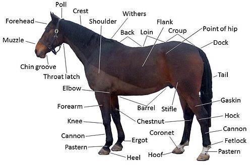
Equine anatomy
Horses and other equids evolved as grazing animals, adapted to eating small amounts of the same kind of food all day long.
In the wild, the horse adapted to eating prairie grasses in semi-arid regions and traveling significant distances each day in order to obtain adequate nutrition.
Horses select pieces of forage and pick up finer foods, such as grain, with their sensitive, prehensile lips.
The front teeth of the horse, called incisors, clip forage, and food is then pushed back in the mouth by the tongue, and ground up for swallowing by the premolars and molars.
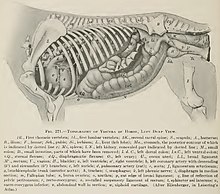
Bile from the liver aids in digesting fats in the duodenum combined with enzymes from the pancreas and small intestine.
The cecum (called the "water gut" in old textbooks) is the first section of the large intestine, analogous to the appendix in humans.
It contains bacteria and other microbes that break down cellulose and other indigestible plant fiber through fermentation into volatile fatty acids.
The large colon continues the fermentation process, and absorbs volatile fatty acids as an energy source.
Due to its many twists and turns, the large colon is a common place for certain forms of colic including impaction, displacement and volvulus.
It is the area where the majority of water in the horse's diet is absorbed, and is the place where fecal balls are formed.
The rectum is about 30 cm (1 ft) long, and acts as a holding chamber for waste matter, which is then expelled from the body via the anus.
[14] The mare's reproductive system is responsible for controlling gestation, birth, and lactation, as well as her estrous cycle and mating behavior.
It lies ventral to the 4th or 5th lumbar vertebrae, although its position within the mare can vary depending on the movement of the intestines and distention of the bladder.
[23] The stallion's reproductive system is responsible for his sexual behavior and secondary sex characteristics (such as a large crest).
A horse's incisors, premolars, and molars, once fully developed, continue to erupt throughout its lifetime as the grinding surface is worn down through chewing.
The hoof of the horse encases part of the second and all of the third phalanx of the lower limbs, analogous to the fingertip or toe tip of a human.
The hoof wall is a much larger, thicker and stronger version of the human fingernail or toenail, made up of similar materials, primarily keratin, a very strong protein molecule.
The horse's hoof contains a high proportion of sulfur-containing amino acids which contribute to its resilience and toughness.
Vascular fold-like structures called laminae suspend the distal phalanx from the hoof wall.
Due to their relatively poor blood supply, ligament injuries generally take a long time to heal.
They are a major contributor to shock absorption, are necessary for support of the horse's body, and translate the force generated by muscles into movement.
The horse's respiratory system consists of the nostrils, pharynx, larynx, trachea, diaphragm, and lungs.
The genus Equus also has a unique part of the respiratory system called the guttural pouch, which is thought to equalize air pressure on the tympanic membrane.
Some of this effect may be lost when a horse is shod (eliminating the expansion and contraction of the hoof wall and raising the frog higher from the ground).