Humerus
The humeral upper extremity consists of a rounded head, a narrow neck, and two short processes (tubercles, sometimes called tuberosities).
[1] The upper or proximal extremity of the humerus consists of the bone's large rounded head joined to the body by a constricted portion called the neck, and two eminences, the greater and lesser tubercles.
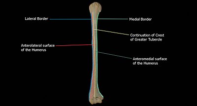
The crest of the greater tubercle forms the lateral lip of the bicipital groove and is the site for insertion of pectoralis major.
The crest of the lesser tubercle forms the medial lip of the bicipital groove and is the site for insertion of teres major and latissimus dorsi muscles.
In the fresh state its upper part is covered with a thin layer of cartilage, lined by a prolongation of the synovial membrane of the shoulder-joint; its lower portion gives insertion to the tendon of the latissimus dorsi muscle.
Its lips are called, respectively, the crests of the greater and lesser tubercles (bicipital ridges), and form the upper parts of the anterior and medial borders of the body of the bone.
The posterorsuperior part of the shaft has a crest, beginning just below the surgical neck of the humerus and extends till the superior tip of the deltoid tuberosity.
The lateral portion of this surface consists of a smooth, rounded eminence, named the capitulum of the humerus; it articulates with the cup-shaped depression on the head of the radius, and is limited to the front and lower part of the bone.
Above the front part of the capitulum is a slight depression, the radial fossa, which receives the anterior border of the head of the radius, when the forearm is flexed.
These fossæ are separated from one another by a thin, transparent lamina of bone, which is sometimes perforated by a supratrochlear foramen; they are lined in the fresh state by the synovial membrane of the elbow-joint, and their margins afford attachment to the anterior and posterior ligaments of this articulation.
The grooved portion of the articular surface fits accurately within the semilunar notch of the ulna; it is broader and deeper on the posterior than on the anterior aspect of the bone, and is inclined obliquely downward and forward toward the medial side.
Signs and symptoms of this dislocation include a loss of the normal shoulder contour and a palpable depression under the acromion.
At the midshaft of the humerus, the radial nerve travels from the posterior to the anterior aspect of the bone in the spiral groove.
[citation needed] The deltoid originates on the lateral third of the clavicle, acromion and the crest of the spine of the scapula.
The infraspinatus and teres minor insert on the greater tubercle, and work to laterally, or externally, rotate the humerus.
In contrast, the subscapularis muscle inserts onto the lesser tubercle and works to medially, or internally, rotate the humerus.
The four muscles of supraspinatus, infraspinatus, teres minor and subscapularis form a musculo-ligamentous girdle called the rotator cuff.
Primitive fossils of amphibians had little, if any, shaft connecting the upper and lower extremities, making their limbs very short.
In many reptiles and some mammals (where it is the primitive state), the lower extremity includes a large opening called the entepicondylar foramen to allow the passage of nerves and blood vessels.
[5] During embryonic development, the humerus is one of the first structures to ossify, beginning with the first ossification center in the shaft of the bone.