Keratoconus
[12] People with early keratoconus often notice a minor blurring or distortion of their vision, as well as an increased sensitivity to light, so they may visit their clinician seeking corrective lenses for reading or driving.
[19] Several sources suggest that keratoconus likely arises from a number of different factors: genetic, environmental or cellular, any of which may form the trigger for the onset of the disease.
As the two come into contact, cellular and structural changes in the cornea adversely affect its integrity and lead to the bulging and scarring characteristic of the disorder.
Scarring appears to be an aspect of the corneal degradation; however, a recent, large, multicenter study suggests abrasion by contact lenses may increase the likelihood of this finding by a factor over two.
[25] Other studies have suggested that reduced activity by the enzyme aldehyde dehydrogenase may be responsible for a build-up of free radicals and oxidising species in the cornea.
[27] While keratoconus is considered a noninflammatory disorder, one study shows wearing rigid contact lenses by people leads to overexpression of proinflammatory cytokines, such as IL-6, TNF-alpha, ICAM-1, and VCAM-1 in the tear fluid.
[31] The frequency of occurrence in close family members is not clearly defined, though it is known to be considerably higher than that in the general population,[19] and studies have obtained estimates ranging between 6% and 19%.
Hormones such as androgen, prolactin, estrogen and progesterone have been shown to influence corneal biomechanics and tissue remodeling, potentially affecting the integrity of the cornea in individuals predisposed to keratoconus.
The eye examination may proceed to measurement of the localized curvature of the cornea with a manual keratometer,[43] with detection of irregular astigmatism suggesting a possibility of keratoconus.
[15][44] If keratoconus is suspected, the ophthalmologist or optometrist will search for other characteristic findings of the disease by means of slit lamp examination of the cornea.
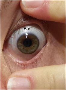
[45] The Fleischer ring, caused by deposition of the iron oxide hemosiderin within the corneal epithelium, is subtle and may not be readily detectable in all cases, but becomes more evident when viewed under a cobalt blue filter.
The topographical map indicates any distortions or scarring in the cornea, with keratoconus revealed by a characteristic steepening of curvature that is usually below the centerline of the eye.
Between 11% and 27% of cases of keratoconus[21][58][59] will progress to a point where vision correction is no longer possible, thinning of the cornea becomes excessive, or scarring as a result of contact lens wear causes problems of its own, and a corneal transplantation or penetrating keratoplasty becomes required.
[62] The long-term outlook for corneal transplants performed for keratoconus is usually favorable once the initial healing period is completed and a few years have elapsed without problems.
[20] The procedure requires a greater level of skill on the part of the surgeon, and is less frequently performed than a penetrating keratoplasty, as the outcome is generally less favorable.
[69] Corneal collagen cross-linking is a developing treatment that aims to strengthen the cornea, however, according to a 2015 Cochrane review, there is insufficient evidence to determine if it is useful in keratoconus.
[72] Radial keratotomy is a refractive surgery procedure where the surgeon makes a spoke-like pattern of incisions into the cornea to modify its shape.
[74][75] Patients with keratoconus typically present initially with mild astigmatism and myopia, commonly at the onset of puberty, and are diagnosed by the late teenage years or early 20s.
[19][76] Patients' vision will seem to fluctuate over a period of months, driving them to change lens prescriptions frequently, but as the condition worsens, contact lenses are required in the majority of cases.
The course of the disorder can be quite variable, with some patients remaining stable for years or indefinitely, while others progress rapidly or experience occasional exacerbations over a long and otherwise steady course.
The patient experiences pain and a sudden severe clouding of vision, with the cornea taking on a translucent milky-white appearance known as a corneal hydrops.
Although a hydrops usually causes increased scarring of the cornea, occasionally it will benefit a patient by creating a flatter cone, aiding the fitting of contact lenses.
[79] Some studies have suggested a higher prevalence amongst females,[82] or that people of South Asian ethnicity are 4.4 times as likely to develop keratoconus as Caucasians, and are also more likely to be affected with the condition earlier.
The German oculist Burchard Mauchart provided an early description in a 1748 doctoral dissertation of a case of keratoconus,[19] which he called staphyloma diaphanum.
[13] In 1859, British surgeon William Bowman used an ophthalmoscope (recently invented by Hermann von Helmholtz) to diagnose keratoconus, and described how to angle the instrument's mirror so as to best see the conical shape of the cornea.
[84] Bowman also attempted to restore vision by pulling on the iris with a fine hook inserted through the cornea and stretching the pupil into a vertical slit, like that of a cat.
He reported that he had had a measure of success with the technique, restoring vision to an 18-year-old woman who had previously been unable to count fingers at a distance of 8 inches (20 cm).
By 1869, when the pioneering Swiss ophthalmologist Johann Horner wrote a thesis entitled On the treatment of keratoconus,[85] the disorder had acquired its current name.
The treatment at that time, endorsed by the leading German ophthalmologist Albrecht von Graefe, was an attempt to physically reshape the cornea by chemical cauterization with a silver nitrate solution and application of a miosis-causing agent with a pressure dressing.
[19] In 1888, the treatment of keratoconus became one of the first practical applications of the then newly invented contact lens, when the French physician Eugène Kalt manufactured a glass scleral shell that improved vision by compressing the cornea into a more regular shape.

"... a candle, when looked at, appears like a number of lights, confusedly running into one another" — Nottingham [ 13 ]