Knee
[2] The knee is a modified hinge joint, which permits flexion and extension as well as slight internal and external rotation.
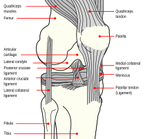
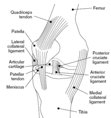
)[5] The knee is a modified hinge joint, a type of synovial joint, which is composed of three functional compartments: the patellofemoral articulation, consisting of the patella, or "kneecap", and the patellar groove on the front of the femur through which it slides; and the medial and lateral tibiofemoral articulations linking the femur, or thigh bone, with the tibia, the main bone of the lower leg.
It plays an essential role in movement related to carrying the body weight in horizontal (running and walking) and vertical (jumping) directions.
Because it is the largest sesamoid bone in the human body, the ossification process takes significantly longer.
[10]: 194–95 The pair of tibial condyles are separated by the intercondylar eminence[10]: 206 composed of a lateral and a medial tubercle.
Between these two extensions, the synovial membrane passes in front of the two cruciate ligaments at the center of the joint, thus forming a pocket direct inward.
[10]: 210 Cartilage is a thin, elastic tissue that protects the bone and makes certain that the joint surfaces can slide easily over each other.
Collagen fibres within the articular cartilage have been described by Benninghoff as arising from the subchondral bone in a radial manner, building so called Gothic arches.
The menisci are flattened at the center of the knee joint, fused with the synovial membrane laterally, and can move over the tibial surface.
[13] The ligaments surrounding the knee joint offer stability by limiting movements and, together with the menisci and several bursae, protect the articular capsule.
[23] This very strong ligament helps give the patella its mechanical leverage[24] and also functions as a cap for the condyles of the femur.
The oblique popliteal ligament is a radiation of the tendon of the semimembranosus on the medial side, from where it is direct laterally and proximally.
The arcuate popliteal ligament originates on the apex of the head of the fibula to stretch proximally, crosses the tendon of the popliteus muscle, and passes into the capsule.
[10]: 206 The most muscles responsible for the movement of the knee joint belong to either the anterior, medial or posterior compartment of the thigh.
The total range of motion is dependent on several parameters such as soft-tissue restraints, active insufficiency, and hamstring tightness.
During extension, the femoral condyles glide and roll into a position which causes the complete unfolding of the tibial collateral ligament.
This terminal rotation is made possible by the shape of the medial femoral condyle, assisted by contraction of the popliteus muscle and the iliotibial tract and is caused by the stretching of the anterior cruciate ligament.
[29] The majority of minor cases of knee pain can be treated at home with rest and ice, but more serious injuries do require surgical care.
The same activity such as climbing stairs may cause pain from patellofemoral compression for someone who is physically unfit, but not for someone else (or even for that person at a different time).
[31] In sports that place great pressure on the knees, especially with twisting forces, it is common to tear one or more ligaments or cartilages.
Minor tears of the anterior cruciate ligament may heal over time, but a torn ACL requires surgery.
Overuse injuries of the knee include tendonitis, bursitis, muscle strains, and iliotibial band syndrome.
[35] Individuals may reduce the chances of overuse injuries by warming up prior to exercise, by limiting high impact activities and keep their weight under control.
[39] Knee osteoarthritis is a major cause of pain and disability worldwide, with prevalence estimated at about 4% of the population, particularly among the elderly.
[40] Radiofrequency ablation of certain knee nerves is an outpatient procedure to reduce chronic arthritic pain.
[14][15][40] Using radiofrequency energy delivered via small electrodes positioned at target genicular nerves, the treatment achieves partial sensory denervation of the joint capsule.
In addition to developing new surgical procedures, ongoing research is looking into underlying problems which may increase the likelihood of an athlete suffering a severe knee injury.
These findings may lead to effective preventive measures, especially in female athletes, who have been shown to be especially vulnerable to ACL tears from relatively minor trauma.
Also in quadrupeds, particularly horses, ungulates, and elephants, the layman's term "knee" also commonly refers to the forward-facing joint in the foreleg, the carpus, which is homologous to the human wrist.
The layman's term "knee" may also refer to the (lower and often more visible due to not being covered by feathers) joint between the tibiotarsus and tarsometatarsus, which is homologous to the human ankle.