Mediastinal shift
[2] Typically, these shifts are observed on x-ray but also on computed tomography (CT) or magnetic resonance imaging (MRI).
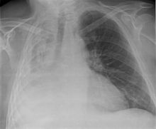
On chest x-ray, tracheal deviation, or movement of the trachea away from its midline position can be used as a sign of a shift.
Tension pneumothorax is an emergent condition in which air gets trapped in the space between the chest wall and the lung.
Forces are transmitted to the mediastinum and effectively "push" the mediastinal structures to the opposite side of the chest.
[3] Hemothorax, or accumulation of blood in the pleural space, can result from trauma or surgical procedures in the chest.
This accumulation of blood can grow large enough to compress the lung and push away other structures in the chest, thus causing a mediastinal shift.
[9] Radiographic appearance is similar to that of a pleural effusion with costophrenic angle blunting and white out of lung zones.
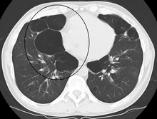
Radiographic features of teratomas typically include fluid and fat but also muscle, teeth, and bones inside the mass.
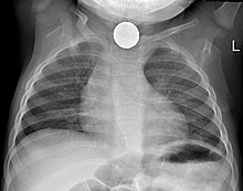
There are numerous etiologies, including post-operative atelectasis, surfactant deficiency, mucus plugging, and foreign body aspiration.
These include agenesis due to gene mutation, fetal hydrothorax, and congenital diaphragmatic hernia.
Notably, patients can experience post pneumonectomy syndrome due to a severe mediastinal shift.
This presents as difficulty breathing due to a shift of airways and rotation of the heart and great vessels.
[20] Foreign body aspiration is a major cause of death in young children due to their underdeveloped swallowing coordination.
The units making up the substructure of the lung (alveoli) become permanently enlarged due to the destruction of their walls.