Medical photography
The nature of the work requires a respect for and sensitivity to people, an awareness of sterile procedures and an adherence to privacy legislation and policies.
This solved a problem of representation by artists who were asked to produce illustrations only from description or highly influenced by the interpretation of physicians and surgeons.
The first application of photography in medicine appears in 1840 when Alfred François Donné of the Charité Hospital in Paris photographed sections of bones and teeth.
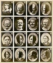
[3] Hugh Welch Diamond, a physician and founding member of the Royal Photographic Society, used photography as a tool in medicine, particularly in the field of mental illness.
With the assistance of Adrien Tournachon, brother of Felix Nadar, he photographed facial expressions and at one point listed 53 emotions that could be identified based on the muscular action.
His work was published in 1862 in Mécanisme de la physionomie humaine in what was the most remarkable of all photographically illustrated books in medical science prior to 1900.
[4] Dr. Jean-Martin Charcot, a student of Duchenne de Boulogne, believed like Diamond that photographs would play a significant role in the diagnosis and management of patients.
Charcot began publishing Nouvelle iconographie de la Salpêtriere in 1888 that used photographs to show clinical presentations of cases at Salpêtrière.
In 1850, Joseph T. Zealy (1812–93) was commissioned by Louis Agassiz to make daguerreotypes of plantation workers of African origin in the southern United States of America.
Thomas Huxley established a system of photographing the human body with fixed views which included a rod of known dimension to make measurements.
Francis Galton believed it was possible to systematically organize traits of inheritable attributes, intellectual, moral and physical with respect to families, groups, classes and racial types.
The Burns Archive Press book Shooting Soldiers: Civil War Medical Photography By Reed B. Bonteco, contains a large selection of these photographs and a history of Bontecou.
[6] Attempts to publish medical photographs in anatomy text books was met with limited success in the early years of photography.
Stereophotography became of interest as a way to add a three-dimensional quality to show the spatial relationships of gross anatomy and clinical case studies.
From 2000, the federal and provincial governments of Canada passed legislation to regulate the use, collection and disclosure of medical photography by healthcare professionals.
As a result, Canadian companies have developed to create specialized mobile apps, such as ShareSmart, and businesses have sought to provide solutions to comply with the new regulatory scheme.