Cataract
[1][4] Risk factors include diabetes, longstanding use of corticosteroid medication, smoking tobacco, prolonged exposure to sunlight, and alcohol.
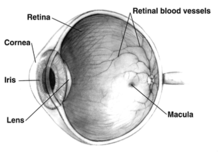
[1] The underlying mechanism involves accumulation of clumps of protein or yellow-brown pigment in the lens that reduces transmission of light to the retina at the back of the eye.
[1] Wearing sunglasses and a wide brimmed hat, eating leafy vegetables and fruits, and avoiding smoking may reduce the risk of developing cataracts, or slow the process.
[14] The severity of cataract formation, assuming no other eye disease is present, is judged primarily by a visual acuity test.
[1][4] Lens proteins denature and degrade over time, and this process is accelerated by diseases such as diabetes mellitus and hypertension.
Environmental factors, including toxins, radiation, and ultraviolet light have cumulative effects which are worsened by the loss of protective and restorative mechanisms due to alterations in gene expression and chemical processes within the eye.
[16] Oxidative stress associated with lipid peroxidation is an important pathogenic mechanism in cataract formation.
[21] Ultraviolet light, specifically UVB, has also been shown to cause cataracts, and some evidence indicates sunglasses worn at an early age can slow its development in later life.
[citation needed] The genetic component is strong in the development of cataracts,[23] most commonly through mechanisms that protect and maintain the lens.
[citation needed] Ichthyosis is an autosomal recessive disorder associated with cuneiform cataracts and nuclear sclerosis.
Although no means of preventing cataracts has been scientifically proven, wearing sunglasses that block ultraviolet light may slow their development.
[57] Extracapsular cataract extraction (ECCE) consists of removing the lens manually, but leaving the majority of the capsule intact.
In MSICS, the lens is removed through a self-sealing scleral tunnel wound in the sclera which, ideally, is watertight and does not require suturing.
[59] The lens and surrounding capsule are removed in one piece through a large incision while pressure is applied to the vitreous membrane.
The patient is usually ambulatory on the day of surgery, but is advised to move cautiously and avoid straining or heavy lifting for about a month.
Retinal detachment frequently presents with unilateral visual field defects, blurring of vision, flashes of light, or floating spots.
Particular risk factors are younger age, male sex, longer axial length, and complications during surgery.
[64] Posterior capsular opacification, also known as after-cataract, is a condition in which months or years after successful cataract surgery, vision deteriorates or problems with glare and light scattering recur, usually due to thickening of the back or posterior capsule surrounding the implanted lens, so-called 'posterior lens capsule opacification'.
The laser can be aimed very accurately, and the small part of the capsule which is cut falls harmlessly to the bottom of the inside of the eye.
This procedure leaves sufficient capsule to hold the lens in place, but removes enough to allow light to pass directly through to the retina.
[65] Posterior capsular opacification is common and occurs following up to one in four operations, but these rates are decreasing following the introduction of modern intraocular lenses together with a better understanding of the causes.
[68] Even where surgical services are available, low vision associated with cataracts may still be prevalent as a result of long waits for, and barriers to, surgery, such as cost, lack of information and transportation problems.
[76] Cataract surgery was first described by the Ayurvedic physician, Suśruta (about 5th century BCE) in Sushruta Samhita in ancient India.
[77] References to cataracts and their treatment in Ancient Rome are also found in 29 AD in De Medicinae, the work of the Latin encyclopedist Aulus Cornelius Celsus.
2nd century CE), a prominent Greek physician, surgeon and philosopher, performed an operation similar to modern cataract surgery.
[80] Muslim ophthalmologist Ammar Al-Mawsili, in his The Book of Choice in Ophthalmology, written circa 1000 CE, wrote of his invention of a syringe and the technique of cataract extraction while experimenting with it on a patient.
[84][85] As rapidly running water turns white, so the term may have been used metaphorically to describe the similar appearance of mature ocular opacities.
[87] Early Persian physicians called the term nazul-i-ah, or "descent of the water"—vulgarised into waterfall disease or cataract—believing such blindness to be caused by an outpouring of corrupt humour into the eye.
The incision at the junction of the sclera and cornea and the hole in capsule during capsulorhexis, traditionally made with a handheld blade, needle, and forceps, are dependent on skill and experience of the surgeon.
[92] Stem cells have been used in a clinical trial, with results submitted in 2014 and published in March 2016, for lens regeneration in twelve children under the age of two with cataracts present at birth.


|
no data
<90
90–180
180–270
270–360
360–450
450–540
|
540–630
630–720
720–810
810–900
900–990
>990
|