Optical coherence tomography
OCT uses coherent near-infrared light to obtain micrometer-level depth resolved images of biological tissue or other scattering media.
The first demonstration of OCT imaging (in vitro) was published by a team from MIT and Harvard Medical School in a 1991 article in the journal Science.
[1] The article introduced the term "OCT" to credit its derivation from optical coherence-domain reflectometry, in which the axial resolution is based on temporal coherence.
[8][9][10][11][12][13][14][15][16][17][18][19][excessive citations] The potential to use interferometry for imaging was proposed,[19] and measurement of retinal elevation profile and thickness had been demonstrated.
[18] The initial commercial clinical OCT systems were based on point-scanning TD-OCT technology, which primarily produced cross-sectional images due to the speed limitation (tens to thousands of axial scans per second).
Over the past three decades, the speed of commercial clinical OCT systems has increased more than 1000-fold, doubling every three years and rivaling Moore's law of computer chip performance.
Development of parallel image acquisition approaches such as line-field and full-field technology may allow the performance improvement trend to continue.
It has greatly improved the management of the top three causes of blindness – macular degeneration, diabetic retinopathy, and glaucoma – thereby preventing vision loss in many patients.
[21] Beyond ophthalmology and cardiology, applications are also developing in other medical specialties such as dermatology, gastroenterology,[22] neurology and neurovascular imaging,[23][24] oncology, and dentistry.
Stemming from single lateral point low-coherence interferometry the addition of a wide range of technologies enabled key milestones in this computational imaging technique.
For their roles in the invention of OCT, Fujimoto, Huang, and Swanson received the 2023 Lasker-DeBakey Clinical Medical Research Award and the National Medal of Technology and Innovation.
Optical coherence tomography (OCT) is a technique for obtaining sub-surface images of translucent or opaque materials at a resolution equivalent to a low-power microscope.
Thus OCT can build up clear 3D images of thick samples by rejecting background signal while collecting light directly reflected from surfaces of interest.
[41] The technique is limited to imaging 1 to 2 mm below the surface in biological tissue, because at greater depths the proportion of light that escapes without scattering is too small to be detected.
is called the complex degree of coherence, i.e. the interference envelope and carrier dependent on reference arm scan or time delay
The axial and lateral resolutions of OCT are decoupled from one another; the former being an equivalent to the coherence length of the light source and the latter being a function of the optics.
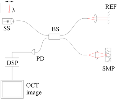
[43] The drawbacks of this technology are found in a strong fall-off of the SNR, which is proportional to the distance from the zero delay and a sinc-type reduction of the depth-dependent sensitivity because of limited detection linewidth.
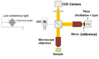
This confocal gate, which is absent in the full-field OCT technique, gives LC-OCT an advantage in terms of detection sensitivity and penetration in highly scattering media such as skin tissues.
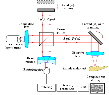
[68] More precisely, interferometric images are created by a Michelson interferometer where the path length difference is varied by a fast electric component (usually a piezo mirror in the reference arm).
The "en-face" tomographic images are thus produced by a wide-field illumination, ensured by the Linnik configuration of the Michelson interferometer where a microscope objective is used in both arms.
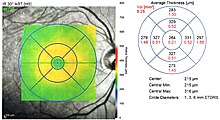
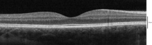
Ocular (or ophthalmic) OCT is used heavily by ophthalmologists and optometrists to obtain high-resolution images of the retina and anterior segment.
Owing to OCT's capability to show cross-sections of tissue layers with micrometer resolution, OCT provides a straightforward method of assessing cellular organization, photoreceptor integrity,[73][74][75][76] and axonal thickness in glaucoma,[77] macular degeneration,[78] diabetic macular edema,[79] multiple sclerosis,[80] optic neuritis,[81] and other eye diseases or systemic pathologies which have ocular signs.
[87] Retinal imaging with PS-OCT demonstrated how the thickness and birefringence of blood vessel wall tissue of healthy subjects could be quantified, in vivo.
[92] In the settings of cardiology, OCT is used to image coronary arteries to visualize vessel wall lumen morphology and microstructure at a resolution ~10 times higher than other existing modalities such as intravascular ultrasounds, and x-ray angiography (intracoronary optical coherence tomography).
Intravascular OCT has been combined with near-infrared fluorescence molecular imaging (NIRF) to enhance its capability to detect molecular/functional and tissue morphological information simultaneously.
[100] Endoscopic/intravascular OCT has been further developed for use in neurovascular applications including imaging for guiding endovascular treatment of ischemic stroke and brain aneurysms.
[115] Emerging high-resolution OCT techniques such as LC-OCT have the potential to improve the clinical diagnostic process, allowing for the early detection of malignant skin tumors – including melanoma – and a reduction in the number of surgical excisions of benign lesions.
[116] Other promising areas of application include the imaging of lesions where excisions are hazardous or impossible and the guidance of surgical interventions through identification of tumor margins.
[129] Due to the high volume of produced pills, an interesting field of application is in the pharmaceutical industry to control the coating of tablets.
Its high-resolution imaging detects defects, characterizes material properties and ensures the integrity of internal geometries without damaging the part.