Orientation column
Orientation columns are organized regions of neurons that are excited by visual line stimuli of varying angles.
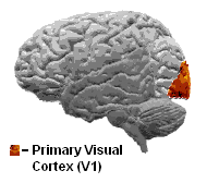
This was found through an experiment by giving a cat specific visual stimuli and measuring the corresponding excitation of the neurons in striate cortex (V1).
The act of changing the slides produced a faint shadow line across the projector, and excited the neuron they were measuring.
His technique used photodiodes to detect optical changes in the visual cortex with the metabolic marker, 2-deoxyglucose, which labels active neurons.
This confirmed Hubel and Wiesel's studies and also brought to light the swirls and pinwheel formations in the striate cortex.
[4] [6] Hubel and Wiesel received the Nobel Prize in Physiology and Medicine in 1981 for their contributions to our knowledge of the development of the visual system.
[10] The second possible advantage is the ordered structure aids in development, by guaranteeing all orientations are represented throughout the visual field with minimal redundancy and no deficiencies.
The third possible advantage is that if columns with similar orientation selectivity are close together, fewer afferents from the LGN are needed.
[12] Also if the visual environment is confined to only vertical or horizontal lines during this critical period the distribution of the preferred orientation of cells in the striate cortex become abnormal.
A highly debated[15][16] model for the origin of orientation maps is Moiré interference from retinal ganglion cells (RGCs).
[17] The ideal case takes two layers of perfect hexagonal lattices of the on-center and off-center receptive fields of the RGCs.
Cortical inputs from this mosaic of RGCs through the LGN can explain the origin of the orientation maps in the visual cortex.
Currently there is research that is testing this hypothesis by "mapping human orientation discrimination thresholds of very small stimuli in the far periphery.