VP1 monomers are generally around 350 amino acids long and are capable of self-assembly into an icosahedral structure consisting of 360 VP1 molecules organized into 72 pentamers.
The VP1 protein, along with capsid components VP2 and VP3, is expressed from the "late region" of the circular viral genome.
The flexibility of the C-terminal arm will enable it to adopt different conformations in the six distinct interaction environments imposed by the symmetry of the icosahedral assembly.
VP1 has a nuclear localization sequence that enables import from the cytoplasm where it is synthesized by the host translation machinery to the cell nucleus where new virions are assembled.
This nuclear import process, mediated by karyopherins, acts on assembled VP1 pentamers in complex with VP2 or VP3; oligomerization to form capsids occurs in the nucleus.
The structure of an individual pentamer of the murine polyomavirus VP1 protein. Each monomer is colored differently. The conformationally flexible C-terminal arms are shown here in conformations compatible with binding to neighboring molecules. Superposed is a fragment of the polyomavirus VP2 protein (white), which binds to a pentamer oriented toward the central cavity. VP1 is from
PDB
:
1SIE
; VP2 is from
PDB
:
1CN3 1CN3
.
The same capsid structure as above, colored to illustrate the assembly of the icosahedral architecture from VP1 pentamers. Each symmetry-related VP1 monomer is shown in a different color. From
PDB
:
1SIE
.
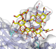
Murine polyomavirus
VP1 in complex with the GT1a
glycan
. GT1a is shown in yellow and the VP1 monomer with a white surface and a blue protein backbone. A complex network of
hydrogen bonds
, many water-mediated, is shown at the binding surface by orange lines, with participating protein residues shown as sticks. Mutations of the two residues shown in cyan at the bottom of the figure can significantly affect pathogenicity. From
PDB
:
5CPW
.
[
3
]