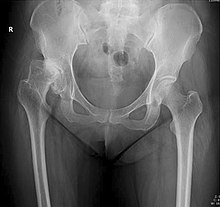
X-ray of hip dysplasia
[1][2] Ultrasound imaging yields better results defining the anatomy until the cartilage is ossified.
Reliability of measurements increases if indicators of pelvic alignment are taken into account: The most useful lines and angles that can be drawn in the pediatric pelvis assessing hip dysplasia are as follows:[3] In the adult hip there are important landmarks to be recognized on plain film radiographs:[3] On CT, the anterior center-edge Lequesne’s angle can be measured in a false profile view of the hip or in a sagittal CT scan.
[3] In 1979 Dr. John F. Crowe et al. proposed a classification to define the degree of malformation and dislocation.
Rather than using the Wiberg angle because it makes it difficult to quantify the degree of dislocation they used 3 key elements to determine the degree of subluxation: A reference line at the lower rim of the "teardrop", junction between the femoral head and neck of the respective joint and the height of the pelvis (vertical measurement).
They studied anteroposterior pelvic x-rays and drew horizontal lines through the lower rim of a feature called "teardrop".