Tibia
The tibia is connected to the fibula by the interosseous membrane of leg, forming a type of fibrous joint called a syndesmosis with very little movement.
As in other vertebrates the tibia is one of two bones in the lower leg, the other being the fibula, and is a component of the knee and ankle joints.
Together with the medial and lateral condyle the intercondylar region forms the tibial plateau, which both articulates with and is anchored to the lower extremity of the femur.
The anterolateral region of the anterior intercondylar area are perforated by numerous small openings for nutrient arteries.
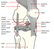
[1] Beneath the condyles is the tibial tuberosity which serves for attachment of the patellar ligament, a continuation of the quadriceps femoris muscle.
Between the articular facets in the intercondylar area, but nearer the posterior than the anterior aspect of the bone, is the intercondyloid eminence (spine of tibia), surmounted on either side by a prominent tubercle, on to the sides of which the articular facets are prolonged; in front of and behind the intercondyloid eminence are rough depressions for the attachment of the anterior and posterior cruciate ligaments and the menisci.
The lateral condyle presents posteriorly a flat articular facet, nearly circular in form, directed downward, backward, and lateralward, for articulation with the head of the fibula.
Just below this a part of the extensor digitorum longus takes origin and a slip from the tendon of the biceps femoris is inserted.
It is sinuous and prominent in the upper two-thirds of its extent, but smooth and rounded below; it gives attachment to the deep fascia of the leg.
From its middle third some fibers of the soleus and flexor digitorum longus muscles take origin.
The medial surface is smooth, convex, and broader above than below; its upper third, directed forward and medialward, is covered by the aponeurosis derived from the tendon of the sartorius, and by the tendons of the Gracilis and Semitendinosus, all of which are inserted nearly as far forward as the anterior crest; in the rest of its extent it is subcutaneous.
The posterior surface presents, at its upper part, a prominent ridge, the popliteal line, which extends obliquely downward from the back part of the articular facet for the fibula to the medial border, at the junction of its upper and middle thirds; it marks the lower limit of the insertion of the Popliteus, serves for the attachment of the fascia covering this muscle, and gives origin to part of the Soleus, Flexor digitorum longus, and Tibialis posterior.
The middle third of the posterior surface is divided by a vertical ridge into two parts; the ridge begins at the popliteal line and is well-marked above, but indistinct below; the medial and broader portion gives origin to the Flexor digitorum longus, the lateral and narrower to part of the Tibialis posterior.
Ossification begins in the center of the body, about the seventh week of fetal life, and gradually extends toward the extremities.
Two additional centers occasionally exist, one for the tongue-shaped process of the upper epiphysis, which forms the tuberosity, and one for the medial malleolus.
Its bending moment in the sagittal plane in the late stance phase is up to 71.6 bodyweight times millimetre.
The tuberosity of the tibia, a crest to which the patellar ligament attaches in mammals, is instead the point for the tendon of the quadriceps muscle in reptiles, birds, and amphibians, which have no patella.