Sphenoid bone
[6] The sphenoid articulates with the frontal, parietal, ethmoid, temporal, zygomatic, palatine, vomer, and occipital bones and helps to connect the neurocranium to the facial skeleton.
It shows: Sphenoidal crest articulates with the perpendicular plate of ethmoid leading to formation of a part of the septum of nose.
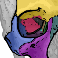
It presents (starting from the front): This is divided into (by infratemporal crest): Foramen pierce it: This forms the posterior wall of the orbit[4] These are two triangular wings projecting laterally from anterosuperior part of the body.
Soon after, the centers for the postsphenoid part of the body appear, one on either side of the sella turcica, and become blended together about the middle of fetal life.
Between the pre- and postsphenoid there are occasionally seen the remains of a canal, the canalis cranio-pharyngeus, through which, in early fetal life, the hypophyseal diverticulum of the buccal ectoderm is transmitted.
The sphenoidal sinuses are present as minute cavities at the time of birth (Onodi), but do not attain their full size until after puberty.
Finally, the basisphenoid bone formed part of the floor of the braincase and lay immediately above the parasphenoid.
[8] Aside from the loss of the flexible joint at the rear of the palate, this primitive pattern is broadly retained in reptiles, albeit with some individual modifications.
The epipterygoids have extended into the wall of the cranium; they are referred to as alisphenoids when separate in mammals, and form the greater wings of the sphenoid when fused into a larger structure.
These bones remain separate and are the: This article incorporates text in the public domain from page 147 of the 20th edition of Gray's Anatomy (1918)