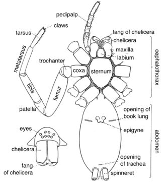
Spider anatomy
These characteristics include bodies divided into two tagmata (sections or segments), eight jointed legs, no wings or antennae, the presence of chelicerae and pedipalps, simple eyes, and an exoskeleton, which is periodically shed.
All spiders are capable of producing silk of various types, which many species use to build webs to ensnare prey.
The exception to this rule are the assassin spiders in the family Archaeidae, whose cephalothorax is divided into two parts by an elongated "neck".
Most external appendages on the spider are attached to the cephalothorax, including the eyes, chelicerae and other mouthparts, pedipalps and legs.
Like other arachnids, spiders are unable to chew their food, so they have a mouth part shaped like a short drinking straw that they use to suck up the liquefied insides of their prey.
Since they do not have antennae, spiders use specialised and sensitive setae on their legs to pick up scent, sounds, vibrations and air currents.
[9] Net-casting spiders of genus Deinopis have their posterior median eyes enlarged into large forward-facing compound lenses.
A fold, known as the epigastric furrow, separates the region of the book lungs and epigyne from the more posterior part of the abdomen.
[12] The abdomen has no appendages except from one to four (usually three) modified pairs of movable telescoping organs called spinnerets, which produce silk.
All other spiders have the spinnerets further towards the posterior end of the body where they form a small cluster, and the anterior central spinnerets on the tenth segment are lost or reduced (suborder Mygalomorphae), or modified into a specialised and flattened plate called the cribellum (suborder Araneomorphae).
The cribellum (usually separated into a left and a right half) produces a thread made up of hundreds to thousands of very fine dry silk fibers (about 10 nm thick) around a few thicker core fibers, which then are combed into a woolly structure by using a group of specialized hairs on their fourth pair of legs.
[13] The cribellate spiders were the first spiders to build specialized prey-catching webs, later evolving into groups that used the spinnerets solely to make webs, instead using silk threads dotted with droplets of a sticky liquid (like pearls on a necklace) to capture small arthropods, and a few large species even small bats and birds.
Rather, their bodies are filled with haemolymph, which is pumped through arteries by a heart into spaces called sinuses surrounding their internal organs.
This is also the case for some basal araneomorph spiders like the family Hypochilidae, but the remaining members of this group have just the anterior pair of book lungs intact while the posterior pair of breathing organs are partly or fully modified into tracheae, through which oxygen is diffused into the haemolymph or directly to the tissue and organs.
Among smaller araneomorph spiders there are species in which the anterior pair of book lungs have also evolved into tracheae, or are simply reduced or missing.
Some very small spiders in moist and sheltered habitats do not have any breathing organs at all, as gas exchange occurs directly through their body surface.
In primitive spiders, such as the Mesothelae and the Mygalomorphae, two pairs of coxal glands open onto the posterior side of the first and third coxae.
Other spiders with more powerfully built chelicerae masticate the entire body of their prey and leave behind only a relatively small amount of indigestible materials.
In 1843 it was revealed that males build a nuptial web into which they deposit a drop of semen, which is then taken up by the copulatory apparatus (the palpal bulb) in the pedipalp.
While the widened palpal tarsus of the southern house spider, Kukulcania hibernalis (Filistatidae), only forms a simple bulb containing the coiled blind duct, members of the genus Argiope have a highly complex structure.