Superficial siderosis
Superficial hemosiderosis of the central nervous system is a disease of the brain resulting from chronic iron deposition in neuronal tissues associated with cerebrospinal fluid.
[6] The excess free iron is circulated in the cerebrospinal fluid and deposited in neuronal tissues, where it catalyzes the formation of reactive oxygen species which can damage DNA, lipids and proteins, and is otherwise toxic to the cells.
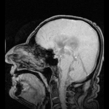
[6] Iron deposition is most prevalent in the inferior temporal lobes, the brainstem, the cerebellum, peri-ventricular structures, the spinal cord, and cranial nerve VIII.
[8] The presence of ‘foamy’, ‘spheroid’, or ‘cytoid’ bodies in affected neuronal tissues has been noted but what they are and their origin remains somewhat unclear.
[8] Taking samples of cerebrospinal fluid may also reveal siderosis through xanthochromia, elevated presence of red blood cells, high iron and ferritin concentrations, and elevated levels of the proteins Tau, amyloid beta (Aβ42), neurofilament light chain (NFL), and glial fibrillary acidic protein (GFAP), but the CSF is sometimes normal.
[10] Detection is complicated by the fact that superficial siderosis is a rare disease and is not well described in neurological texts, so it may go unnoticed until noticeable symptoms appear.
[12] Deferiprone use is associated with severe neutropenia, but this risk and associated complications are usually minimised by administering a weekly blood test that measures absolute neutrophil count.