Thoracic vertebrae
By convention, the human thoracic vertebrae are numbered T1–T12, with the first one (T1) located closest to the skull and the others going down the spine toward the lumbar region.
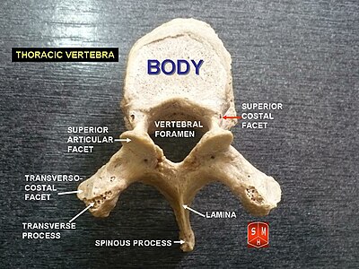
They present, on either side, two costal demi-facets, one above, near the root of the pedicle, the other below, in front of the inferior vertebral notch; these are covered with cartilage in the fresh state, and, when the vertebrae are articulated with one another, form, with the intervening intervertebral fibrocartilages, oval surfaces for the reception of the heads of the ribs.
The laminae are broad, thick, and imbricated – that is to say, they overlap those of subjacent vertebrae like tiles on a roof and connect with the pedicles to surround and protect the spinal cord.
The spinous process is long, triangular on coronal section, directed obliquely downward, arising from the lamina and ending in a tuberculated extremity.
The transverse processes arise from the arch behind the superior articular processes and pedicles; they are thick, strong, and of considerable length, directed obliquely backward and lateralward, and each ends in a clubbed extremity, on the front of which is a small, concave surface, for articulation with the tubercle of a rib.
The superior articular surfaces are directed upward and backward; the spinous process is thick, long, and almost horizontal.
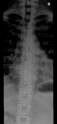
The human trachea divides into two main bronchi at the level of the 5th thoracic vertebra, but may also end higher or lower, depending on breathing.
The tenth thoracic vertebra has an entire articular facet (not demi-facet) on either side, which is placed partly on the lateral surface of the pedicle.
The articular facets for the heads of the ribs are of medium size, and placed chiefly on the pedicles, which are thicker and stronger in this and the next vertebra than in any other part of the thoracic region.
Traces of similar elevations are found on the transverse processes of the tenth and eleventh thoracic vertebrae.