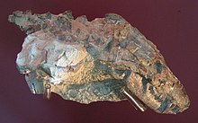
Thrinaxodon
Thrinaxodon is an extinct genus of cynodonts which lived in what are now South Africa and Antarctica during the Late Permian - Early Triassic.
Their unique secondary palate successfully separated the nasal passages from the rest of the mouth, allowing the Thrinaxodon to continue mastication without interrupting to breathe, an adaptation important for digestion.
[3] The arrangement of foramina on the snout of Thrinaxodon resembles that of lizards, such as Tupinambis, and also bears a single large infraorbital foramen.
This adaptation would have allowed the Thrinaxodon to mash its food to a greater extent, decreasing the amount of time necessary for digestion.
The large palatal roof component of the vomer in Thrinaxodon is just dorsal to the choana, or interior nasal passages.
The pterygoid bones extend in the upper jaw and enclose small interpterygoid vacuities that are present on each side of the cultriform processes of the parasphenoids.
Such smooth muscle interactions have been interpreted to be indicative of the tympanum and give the implications that this recess, in conjunction with the fenestra ovalis, outline the origin of the ear in Thrinaxodon.
The most notable jump in bone number reduction had occurred between Thrinaxodon and Probainognathus, a change so dramatic that it is most likely that the fossil record for this particular transition is incomplete.
The canines of T. liorhinus possess small dorsoventrally-directed facets on their surfaces, which appear to be involved with occlusion (dentition alignment in upper- and lower jaw closure).
Some older specimens have been found that possess no multiple-cups lower canines, possibly a response to old age or teeth replacement.
Each of the bones contains a large abundance of globular osteocyte lacunae which radiate a multitude of branched canaliculi.
Combine this with the greater organization of osteocyte lacunae in the periphery of adult T. liorhinus, and we approach the assumption that this creature grew very quickly in order to reach adulthood at an accelerated rate.
The remainder of the femur is made of fibro-lamellar tissue; however, the globular osteocyte lacunae become much more organized and the primary osteons assume less vasculature than many other bones as you begin to approach the subperiosteal surface.
The data actually showed a slight difference between the two, in that the African T. liorhinus contained 26 presacrals, while the Antarctic Thrinaxodon had 27 pre-sacrals.
This discovery was one of many to support the idea of a connected land mass, and that during the early Triassic, Africa and Antarctica must have been linked in some way, shape or form.
Much of the data assumes that the length of the sagittal crest increased at a greater rate in relation to the rest of the skull.
In cynodonts such as Thrinaxodon, the distal femoral condyle articulates with the acetabulum in a way that permits the hindlimb to present itself at a 45-degree angle to the rest of the system.
This is a large difference in comparison to the distal femoral condyle of pelycosaurs, which permits the femur to be parallel with the ground, forcing them to assume a sprawling-like posture.
[2] More interesting is that there is an adaptation that has only been observed within Thrinaxodontidae, which allows them to assume upright posture, similar to that of early Mammalia, within their burrows.
The thoracic segment of the vertebrae contain ribs with large intercostal plates that most likely assisted with either protection or supporting the main frame of the back.
Due to the evolution of a segmented vertebral column into thoracic, lumbar and sacral vertebrae, Thrinaxodon was able to achieve flexibilities that permitted it to comfortably rest within smaller burrows, which may have led to habits such as aestivation or torpor.
This evolution of a segmented rib cage suggests that this may have been the first instance of a diaphragm in the synapsid fossil record; however, without the proper soft tissue impressions this is nothing more than an assumption.
[13][1] The earliest discovery of a burrowing Thrinaxodon places the specimen found around 251 million years ago, a time frame surrounding the Permian–Triassic extinction event.
This behavior had been seen at a relatively low occurrence in the pre-Cenozoic, dominated by therapsids, early-Triassic cynodonts and some early Mammalia.
The changes in vertebral/rib anatomy that arose in Thrinaxodon permit the animals to a greater range of flexibility, and the ability to place their snout underneath their hindlimbs, an adaptive response to small living quarters, in order to preserve warmth and/or for aestivation purposes.
The synchrotron revealed an injured rhinesuchid, Broomistega putterilli, showing signs of broken or damaged limbs and two skull perforations, most likely inflicted by the canines of another carnivore.