Ultrafast laser spectroscopy
Most measurements are done by employing a sequence of ultrashort light pulses to initiate a process and record its dynamics.
Typical Ti:sapphire oscillator pulses have nJ energy and repetition rates 70-100 MHz.
The wide tunability range, high output power, and pulsed or CW operation make the dye laser particularly useful in many physical & chemical studies.
The pump light from the laser diode will excite a state in the doped fiber which can then drop in energy causing a specific wavelength to be emitted.
The strong Coulomb force due to the ionized material in the center of the cloud quickly accelerates the electrons back to the nuclei left behind.
Methods allowing for complete characterization of pulses include frequency-resolved optical gating (FROG) and spectral phase interferometry for direct electric-field reconstruction (SPIDER).
Pulse shapers usually refer to optical modulators which apply Fourier transforms to a laser beam.
It was first observed in 1987 by McPherson et al. who successfully generated harmonic emission up to the 17th order at 248 nm in neon gas.
It is realizable on a laboratory scale (table-top systems) as opposed to large free electron-laser facilities.
High harmonic generation in atoms is well understood in terms of the three-step model (ionization, propagation, and recombination).
Ionization: The intense laser field modifies the Coulomb potential of the atom, electron tunnels through the barrier and ionize.
Recombination: When the field reverses, the electron is accelerated back toward the ionic parent and releases a photon with very high energy.
For this reason, frequency conversion techniques are commonly used to extend the operational spectrum of existing laser light sources.
The most widespread conversion techniques rely on using crystals with second-order non-linearity to perform either parametric amplification or frequency mixing.
A probing light source, typically a xenon arc lamp or broadband laser pulse created by supercontinuum generation, is used to obtain an absorption spectrum of the compound at various times following its excitation.
After passing through the sample, the unabsorbed probe light continues to a photodetector such as an avalanche photodiode array or CMOS camera, and the data is processed to generate an absorption spectrum of the excited state.
Since all the molecules or excitation sites in the sample will not undergo the same dynamics simultaneously, this experiment must be carried out many times (where each "experiment" comes from a single pair of pump and probe laser pulse interactions), and the data must be averaged to generate spectra with accurate intensities and peaks.
The pulsed laser in this setup is used both as a primary excitation source, and a clock signal for the ultrafast measurements.
Although laborious and time-consuming, the monochromator position may also be shifted to allow absorbance decay profiles to be constructed, ultimately to the same effect as the above method.
The data of UTA measurements usually are reconstructed absorption spectra sequenced over the delay time between the pump and probe.
Different frequencies can probe various dynamic molecular processes to differentiate between inhomogeneous and homogeneous line broadening as well as identify coupling between the measured spectroscopic transitions.
2D spectroscopy using ultrafast pulses can be combined with complementary experimental methods to characterize the system under study.
[14] Another technique called Serial Time-encoded amplified microscopy has shown to have the capability of even earlier detection of trace amounts of cancer cells in the blood.
[17][9] Examples of these include the cis-trans photoisomerization of the rhodopsin chromophore retinal, excited state and population dynamics of DNA, and the charge transfer processes in photosynthetic reaction centers[9] Charge transfer dynamics in photosynthetic reaction centers has a direct bearing on man’s ability to develop light harvesting technology, while the excited state dynamics of DNA has implications in diseases such as skin cancer.
The possibility of using a femtosecond technique to study bimolecular reactions at the individual collision level is complicated by the difficulties of spatial and temporal synchronization.
One way to overcome this problem is through the use of Van der Waals complexes of weakly bound molecular cluster.
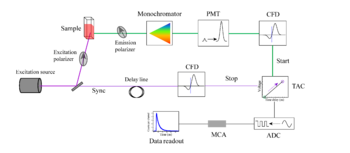
In other words, less than one emitted photon is detected per laser pulse, and the process is repeated multiple times to get an average value.
The fluorescence decay curve is obtained by plotting the measured time on the x-axis and the number of photons detected on the y-axis.
The IRF, which represents the shortest time profile the instrument can detect, serves as a reference for accurately deconvolving the measured data.
Though non-linear least squares analysis can usually detect the different rate constants, determining the processes involved is often very difficult and requires the combination of multiple ultra-fast techniques.