Uterus
: uteri or uteruses) or womb (/wuːm/) is the organ in the reproductive system of most female mammals, including humans, that accommodates the embryonic and fetal development of one or more fertilized eggs until birth.
[1] The uterus is a hormone-responsive sex organ that contains glands in its lining that secrete uterine milk for embryonic nourishment.
In the human, the lower end of the uterus is a narrow part known as the isthmus that connects to the cervix, the anterior gateway leading to the vagina.
It will have divided on its journey to form a blastocyst that will implant itself into the lining of the uterus – the endometrium, where it will receive nutrients and develop into the embryo proper, and later fetus, for the duration of the pregnancy.
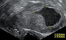
In humans, the uterus is located within the pelvic region immediately behind and almost overlying the bladder, and in front of the sigmoid colon.
During pregnancy, the uterine glands and blood vessels in the endometrium further increase in size and number and form the decidua.
Vascular spaces fuse and become interconnected, forming the placenta, which supplies oxygen and nutrition to the embryo and fetus.
Under normal circumstances, the suspensory part keeps the uterus in anteflexion and anteversion (in 90% of women) and keeps it "floating" in the pelvis.
In males, anti-Müllerian hormone (AMH) secreted from the testes leads to the ducts' regression.
In humans, the lower segments of the two ducts fuse to form a single uterus; in cases of uterine malformations this fusion may be disturbed.
The different uterine morphologies among the mammals are due to varying degrees of fusion of the Müllerian ducts.
[23] When normal labor begins, the uterus forcefully contracts as the cervix dilates, which results in delivery of the infant.
[22] The withdrawal of female sex hormones, estrogen and progesterone, which occurs in the absence of fertilization, triggers the shedding of the functional layer of the endometrium.
A hysterectomy is the surgical removal of the uterus, which may be carried out for a number of reasons including the ridding of tumours both benign and malignant.
A partial hysterectomy may just involve the removal of the uterine body while leaving the cervix intact.
[29][30][31] Most animals that lay eggs, such as birds and reptiles, including most ovoviviparous species, have an oviduct instead of a uterus.
[33][34] Marsupial embryos form a choriovitelline placenta (which can be thought of as something between a monotreme egg and a "true" placenta), in which the egg's yolk sac supplies a large part of the embryo's nutrition but also attaches to the uterine wall and takes nutrients from the mother's bloodstream.
In monotremes such as the platypus, the uterus is duplex and rather than nurturing the embryo, secretes the shell around the egg.
[36] Two uteri usually form initially in a female and usually male fetus, and in placental mammals, they may partially or completely fuse into a single uterus depending on the species.


2. Anteversion with marked anteflexion
3. Anteversion with retrocession
4. Retroversion
5. Retroversion with retroflexion