Uveitis
[1] The uvea consists of the middle layer of pigmented vascular structures of the eye and includes the iris, ciliary body, and choroid.
Treatment typically involves the use of topical eye drop steroids, intravitreal injection, newer biologics, and treating any underlying disease.
While initial treatment is usually successful, complications include other ocular disorders, such as uveitic glaucoma, retinal detachment, optic nerve damage, cataracts, and in some cases, a permanent loss of vision.
Uveitis is classified anatomically into anterior, intermediate, posterior, and panuveitis forms—based on the part of the eye primarily affected.
HLA-B27 AAU has characteristic clinical features including male preponderance, unilateral alternating acute onset, a non-granulomatous appearance, and frequent recurrences, whereas HLA-B27 negative AAU has an equivalent male to female onset, bilateral chronic course, and more frequent granulomatous appearance.
Uveitis is driven by the Th17 T cell sub-population that bear T-cell receptors specific for proteins found in the eye.
[16] These are often not deleted centrally whether due to ocular antigen not being presented in the thymus (therefore not negatively selected) or a state of anergy is induced to prevent self targeting.
The cause of non-infectious uveitis is unknown but there are some strong genetic factors that predispose disease onset including HLA-B27[22][23] and the PTPN22 genotype.
[24] Recent evidence has pointed to reactivation of herpes simplex, varicella zoster and other viruses as important causes of developing what was previously described as idiopathic anterior uveitis.
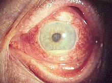
[1] Diagnosis includes dilated fundus examination to rule out posterior uveitis, which presents with white spots across the retina along with retinitis and vasculitis.
Uveitis is typically treated with glucocorticoid steroids, either as topical eye drops (prednisolone acetate) or as oral therapy.
[30] Intravitrial injection of steroid has proven to be a newer useful way to control inflammation for longer without the need for daily eyedrops.
[33] In the case of herpetic uveitis, anti-viral medications, such as valaciclovir or aciclovir, may be administered to treat the causative viral infection.
[36] For non-infectious uveitis, women are more likely (57%) to be affected than men, possibly due to their higher prevalence of related autoimmune diseases.
[37] The prognosis is generally good for those who receive prompt diagnosis and treatment, but serious complication including cataracts, uveitic glaucoma, band keratopathy, macular edema and permanent vision loss may result if left untreated.