Vestibulo-ocular reflex
The reflex acts to stabilize images on the retinas of the eye during head movement.
Since slight head movement is present all the time, VOR is necessary for stabilizing vision: people with an impaired reflex find it difficult to read using print, because the eyes do not stabilise during small head tremors, and also because damage to reflex can cause nystagmus.
It can also be activated by hot or cold stimulation of the inner ear, where the vestibular system sits, and works even in total darkness or when the eyes are closed.
Humans have semicircular canals, neck muscle "stretch" receptors, and the utricle (gravity organ).
Though the semicircular canals cause most of the reflexes which are responsive to acceleration, the maintaining of balance is mediated by the stretch of neck muscles and the pull of gravity on the utricle (otolith organ) of the inner ear.
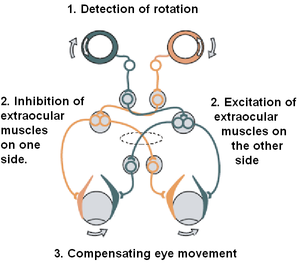
One pathway projects directly to the lateral rectus muscle of the eye via the abducens nerve.
Another nerve tract projects from the abducens nucleus by the medial longitudinal fasciculus to the oculomotor nucleus of the opposite side, which contains motor neurons that drive eye muscle activity, specifically activating the medial rectus muscle of the eye through the oculomotor nerve.
Another pathway (not in picture) directly projects from the vestibular nucleus through the ascending tract of Deiter's to the medial rectus muscle motor neuron of the same side.
In addition to these direct pathways, which drive the velocity of eye rotation, there is an indirect pathway that builds up the position signal needed to prevent the eye from rolling back to center when the head stops moving.
David A. Robinson discovered that the eye muscles require this dual velocity-position drive, and also proposed that it must arise in the brain by mathematically integrating the velocity signal and then sending the resulting position signal to the motoneurons.
Robinson was correct: the 'neural integrator' for horizontal eye position was found in the nucleus prepositus hypoglossi[8] in the medulla, and the neural integrator for vertical and torsional eye positions was found in the interstitial nucleus of Cajal[9] in the midbrain.
The integrator is leaky, with a characteristic leaking time of 20 s. For example, when the subject is sitting still and focusing on an object, and suddenly the light is turned off, the eyes would return to their neutral position in around 40 seconds even as the subject is attempting to keep the focus.
Research indicates that there exists mechanisms in the brain to suppress the VOR using the active visual (retinal) feedback obtained by watching the object in motion.
[14] In the absence of visual feedback, such as when the object passes behind an opaque barrier, humans can continue to visually track the apparent position of the object using anticipatory (extra-retinal) systems within the brain, and the VOR is also suppressed during this activity.
The VOR can even be cognitively suppressed, such as when following an imagined target with the eyes and head together, although the effect tends to be less dramatic than with visual feedback.
Under such conditions, motor learning adjusts the gain of the VOR to produce more accurate eye motion.
Nearsighted people who habitually wear negative spectacles have lower VOR gain.
Farsighted people or aphakes who habitually wear positive spectacle have higher VOR gain.
People who habitually wear contact lens show no change in VOR gain.
The hypothesis is tested by using an specially patterned optokinetic drum that simulates the visual effect of having a very leaky oculomotor integrator.
After 1 hour of viewing, the integrator becomes "anti-leaky", meaning that its value grows exponentially even in the absence of input.
The unusual vestibular stimulation also caused motion sickness symptoms: illusions of bodily rotations, dizziness, and nausea.
After ethanol is fully metabolized, the cupula returns to normal density first, creating nystagmus in the opposite direction (PAN II) during the hangover.
When the function of the right balance system is reduced, by a disease or by an accident, a quick head movement to the right cannot be sensed properly anymore.
The head impulse test can be done at the bed side and used as a screening tool for problems with a person's vestibular system.
In this diagnostic test, a person wears highly sensitive goggles that detect rapid changes in eye movement.
[25] Another way of testing the VOR response is a caloric reflex test, which is an attempt to induce nystagmus (compensatory eye movement in the absence of head motion) by pouring cold or warm water into the ear.
[27] Summary: Cervico-ocular reflex, also known by its acronym COR, involves the achievement of stabilization of a visual target,[28] and image on the retina, through adjustments of gaze impacted by neck and, or head movements or rotations.