Wrist arthroscopy
[1] There are several therapeutic wrist arthroscopy indications, in this article the focus will be on the TFCC-lesion, the SL-lesion, the dorsal ganglion resection and the distal radius fracture.
The Triangular Fibrocartilage Complex (TFCC) is a fibrous structure covering both the radiocarpal and distal radioulnar joint.
Tears in this ligament occur commonly after a person falls or secondary to a wrist fracture.
Abnormalities in the TFCC are classified with the Palmer Classification, which divides the tears in a traumatic or degenerative stage.
[5] The initial treatment of patients with a suspected tear of the scapholunate and lunotriquetral interosseous ligament is a splint of the wrist.
If the pain and instability persists, one could undergo an open surgery to reconstruct the scapholunate ligament.
Arthroscopy is until today in an experimental stage but research suggest that in the near future it will be a reasonable alternative for open surgery due to faster recovery time.
[6] For a tear in the lunotriquetral ligament, arthroscopic debridement is the prime treatment with a loss or reduction of symptoms of 78-100%.
[7] 28 to 58% of the dorsal ganglia resolve spontaneously, still some patients choose to undergo cosmetic intervention for resection of the ganglion when non-operative treatment failed.
Some examples of this non-operative treatment are immobilization through a splint or aspiration of the ganglion with or without injection of a steroid.
In some cases the ganglia are associated with serious loss of wrist function or weakness in the affected finger, which makes a surgical intervention highly indicated.
Arthroscopic intervention has the advantages of smaller incisions, faster recovery of wrist function and less pain postoperative.
[9] Distal radius fractures might occur when a person falls on an outstretched hand (FOOSH).
Conservative treatment consists of wrist immobilization, oral NSAIDs and/or injection with corticoids.
[12] However, relying on arthroscopic findings in the setting of an unclear preoperative diagnosis yields limited diagnostic benefit.
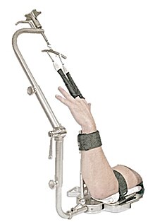
To properly stabilize the wrist, the patient's elbow is flexed and the forearm is immobilized by using a traction apparatus.
Complications of using an irrigation system is that fluid extravasation into the soft tissues of the forearm can result in a compartment syndrome.
[14] To get a clear view of the wrist a high quality camera with a small diameter between 2 and 3 mm is needed.
All probes bear markings at 5 mm intervals to indicate the scale, since the cameras used in arthroscopy have different magnifications.
Every different arthroscopic intervention has its own set of tools, depending on difficulty, type of tissue and available working space.


