Xanthoria parietina
[5] The outer "skin" of the lichen, the cortex, is composed of closely packed fungal hyphae and serves to protect the thallus from water loss due to evaporation as well as harmful effects of high levels of irradiation.
Thalli are much thinner in shady locations than in those exposed to full sunshine; this has the effect of protecting the algae that cannot tolerate high light intensities.
[10] A large number of lichens disperse very effectively by means of symbiotic vegetative propagules such as soredia, isidia and blastidia, and thallus fragmentation.
Two oribatid mite species, Trhypochtonius tectorum and Trichoribates trimaculatus, which are common inhabitants and consumers of X. parietina, are vectors of the photobiont cells.
[5] The increases in nitrate deposition as a result of industrial and agricultural developments in southern Ontario, Canada in the 20th century are thought to be responsible for the reappearance of this species in the local lichen flora.
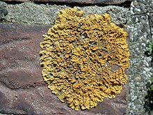
[22][23] Xanthoria parietina produces an orange colored anthraquinone pigment, parietin, that is deposited as tiny crystals in the top layer of the upper cortex.
[26] The biochemical impacts of the lichenicolous fungus Xanthoriicola physciae on its host Xanthoria parietina were investigated using Raman spectroscopy.
This technique revealed that the fungus destroys key photoprotective pigments such as parietin and carotenoids in the host, which are vital for protecting the lichen from intense sunlight.
[27] The water extract of X. parietina has good antiviral activity in vitro, inhibiting the replication of human parainfluenza virus type 2.