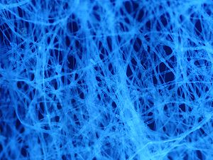
Autofluorescence
[1] The most commonly observed autofluorescencing molecules are NADPH and flavins; the extracellular matrix can also contribute to autofluorescence because of the intrinsic properties of collagen and elastin.
[1] Generally, proteins containing an increased amount of the amino acids tryptophan, tyrosine, and phenylalanine show some degree of autofluorescence.
Light-emitting stains (such as fluorescently labelled antibodies) are applied to samples to enable visualisation of specific structures.
In a few cases, autofluorescence may actually illuminate the structures of interest, or serve as a useful diagnostic indicator.
[6] The super resolution microscopy SPDM revealed autofluorescent cellular objects which are not detectable under conventional fluorescence imaging conditions.