Bioactive glass
The biocompatibility and bioactivity of these glasses has led them to be used as implant devices in the human body to repair and replace diseased or damaged bones.
Hench began development by submitting a proposal hypothesis to the United States Army Medial Research and Development command in 1968 based upon his theory of the body rejecting metallic or polymeric material unless it was able to form a coating of hydroxyapatite which is found in bone.
[5] The glass was batched, melted, and cast into small rectangular implants to be inserted into the femoral bone of rats for six weeks as developed by Dr. Ted Greenlee of the University of Florida.
The 29Si MAS NMR spectroscopy showed that Bioglass 45S5 was a Q2 type-structure with a small amount of Q3; i.e., silicate chains with a few crosslinks.
The 31P MAS NMR revealed predominately Q0 species; i.e., PO43−; subsequent MAS NMR spectroscopy measurements have shown that Si-O-P bonds are below detectable levels [9] There have been many variations on the original composition which was Food and Drug Administration (FDA) approved and termed Bioglass.
High bioactivity is the main advantage of Bioglass, while its disadvantages includes mechanical weakness, low fracture resistance due to amorphous 2-dimensional glass network.
Bioglass can be also used as a bioactive component in composite materials or as powder and can be used to create an artificial septum to treat perforations caused by cocaine abuse.
[11] The first successful surgical use of Bioglass 45S5 was in replacement of ossicles in middle ear, as a treatment of conductive hearing loss.
Annealing is a crucial step in forming bulk parts, due to high thermal expansion of the material.
Heat treatment of Bioglass reduces the volatile alkali metal oxide content and precipitates apatite crystals in the glass matrix.
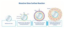
During this activation period, the bioactive glass goes through a series of chemical reactions, creating the ideal conditions for the bone to rebuild through osteoconduction.
Once the hydroxyapatite layer is formed, the bioactive glass interacts with biological entities, i.e., blood proteins, growth factors and collagen.
The bacterial growth inhibiting properties of S53P4 derive from two simultaneous chemical and physical processes, which occurs once the bioactive glass reacts with body fluids.
Sodium (Na) is released from the surface of the bioactive glass and induces an increase in pH (alkaline environment), which is not favorable for the bacteria, thus inhibiting their growth.
The released Na, Ca, Si and P ions give rise to an increase in osmotic pressure due to an elevation in salt concentration, i.e., an environment where bacteria cannot grow.
During the second phase, the Si-O-Si bonds in the silica matrix undergo hydrolysis, yielding a gel-like surface layer rich on Si-O-H groups.
It is commercially available from Mo-Sci Corp. or can be directly prepared by melting a mixture of Na2CO3, K2CO3, MgCO3, CaCO3, SiO2 and NaH2PO4 · 2H2O in a platinum crucible at 1300 °C and quenching between stainless steel plates.
The decrease in the mechanical properties was attributed to the partial conversion of the glass filaments in the scaffolds into a layer mainly composed of a porous hydroxyapatite-like material.
The in vitro study using SBF resulted in a decrease in the compressive strength but the final value was similar to that of human trabecular bone.
[23][24] 13-93 porous glass scaffolds were synthesized using a polyurethane foam replication method in the report by Fu et al.
The resultant curve demonstrated a progressive breaking down of the scaffold structure and the average compressive strength of 11 ± 1 MPa, which was in the range of human trabecular bone and higher than competitive bioactive materials for bone repairing such as hydroxyapatite scaffolds with the same extent of pores and polymer-ceramic composites prepared by the thermally induced phase separation (TIPS) method.
[20] Bioactive glasses have been synthesized through methods such as conventional melting, quenching, the sol–gel process, flame synthesis, and microwave irradiation.
The reasoning behind the introduction of the metallic base is to create a less brittle, stronger material that will be permanently implanted within the body.
Experimentation has been done with sintering double layered, silica-based bioactive glass onto stainless steel substrates at 600 °C for 5 hours.
The underlying mechanisms that enable bioactive glasses to act as materials for bone repair have been investigated since the first work of Hench et al. at the University of Florida.
The inherent porosity of the sol-gel-derived material was cited as a possible explanation for why bioactivity was retained, and often enhanced with respect to the melt-derived glass.
Previously, it was known that a complex interplay existed between bioactive glasses and the molecular biology of the implant host, but the available tools did not provide a sufficient quantity of information to develop a holistic picture.
The first microarray studies on bioactive glasses demonstrated that genes associated with osteoblast growth and differentiation, maintenance of extracellular matrix, and promotion of cell-cell and cell-matrix adhesion were up-regulated by conditioned cell culture media containing the dissolution products of bioactive glass.
S53P4 bioactive glass was first used in a clinical setting as an alternative to bone or cartilage grafts in facial reconstruction surgery.
[30] The use of artificial materials as bone prosthesis had the advantage of being much more versatile than traditional autotransplants, as well as having fewer postoperative side effects.