Tissue (biology)
In biology, tissue is an assembly of similar cells and their extracellular matrix from the same embryonic origin that together carry out a specific function.
Developments in electron microscopy, immunofluorescence, and the use of frozen tissue-sections have enhanced the detail that can be observed in tissues.
With these tools, the classical appearances of tissues can be examined in health and disease, enabling considerable refinement of medical diagnosis and prognosis.
In aquatic plants, aerenchyma tissues, or large air cavities, give support to float on water by making them buoyant.
Spindle shaped fibers are also present in this cell to support them and known as prosenchyma, succulent parenchyma also noted.
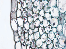
This tissue gives tensile strength to the plant and the cells are compactly arranged and have very little inter-cellular spaces.
These cells have hard and extremely thick secondary walls due to uniform distribution and high secretion of lignin and have a function of providing mechanical support.
The outer epidermis is coated with a waxy thick layer called cutin which prevents loss of water.
The complex permanent tissue consists of more than one type of cells having a common origin which work together as a unit.
Complex tissues are mainly concerned with the transportation of mineral nutrients, organic solutes (food materials), and water.
The common types of complex permanent tissue are: Xylem and phloem together form vascular bundles.
Xylem (Greek, xylos = wood) serves as a chief conducting tissue of vascular plants.
Longer tubes made up of individual cellssels tracheids, while vessel members are open at each end.
[citation needed] Rays are horizontal rows of long-living parenchyma cells that arise out of the vascular cambium.
In spite of the fact that their cytoplasm is actively involved in the conduction of food materials, sieve-tube members do not have nuclei at maturity.
It is the companion cells that are nestled between sieve-tube members that function in some manner bringing about the conduction of food.
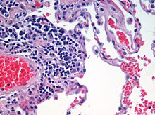
Animal tissues are grouped into four basic types: connective, muscle, nervous, and epithelial.
[4] Collections of tissues joined in units to serve a common function compose organs.
This tissue covers all organismal surfaces that come in contact with the external environment such as the skin, the airways, and the digestive tract.
It serves functions of protection, secretion, and absorption, and is separated from other tissues below by a basal lamina.
The cells comprising an epithelial layer are linked via semi-permeable, tight junctions; hence, this tissue provides a barrier between the external environment and the organ it covers.
Epithelial tissue helps to protect organs from microorganisms, injury, and fluid loss.
Some common kinds of epithelium are listed below: Connective tissues are made up of cells separated by non-living material, which is called an extracellular matrix.
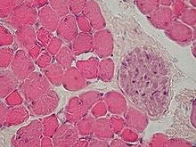
Muscle tissue functions to produce force and cause motion, either locomotion or movement within internal organs.
The filaments are staggered and this is the type of muscle found in earthworms that can extend slowly or make rapid contractions.
[5] In higher animals striated muscles occur in bundles attached to bone to provide movement and are often arranged in antagonistic sets.
Smooth muscle is found in the walls of the uterus, bladder, intestines, stomach, oesophagus, respiratory airways, and blood vessels.
[7] Although he worked without a microscope, Bichat distinguished 21 types of elementary tissues from which the organs of the human body are composed,[8] a number later reduced by other authors.