Cardiac pacemaker
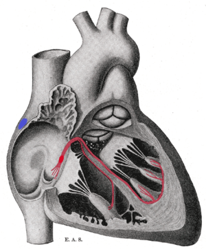
In most humans, these cells are concentrated in the sinoatrial (SA) node, the primary pacemaker, which regulates the heart’s sinus rhythm.
Sometimes a secondary pacemaker sets the pace, if the SA node is damaged or if the electrical conduction system of the heart has problems.
It is a region of cardiac muscle on the wall of the upper right atrium near to the superior vena cava entrance.
Having cardiomyocytes connected via gap junctions allow all contractile cells of the heart to act in a coordinated fashion and contract as a unit.
Cells in the SA node spontaneously depolarize, ultimately resulting in contraction, approximately 100 times per minute.
The cells of the AV node normally discharge at about 40–60 beats per minute, and are called the secondary pacemaker.
The left and right bundle branches, and the Purkinje fibers, will also produce a spontaneous action potential at a rate of 30–40 beats per minute, so if the SA and AV node both fail to function, these cells can become pacemakers.
The SA and AV node do not have fast sodium channels like neurons, and the depolarization is mainly caused by a slow influx of calcium ions.
It is important to note that intracellular calcium causes muscular contraction in contractile cells, and is the effector ion.
For this reason, the pacemaker action potential rising phase slope is more gradual than that of the contractile cell (image 2).
Another important note at this phase is that ionic pumps restore ion concentrations to pre-action potential status.
If the AV node also fails, Purkinje fibers are occasionally capable of acting as the default or "escape" pacemaker.