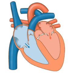
Heart valve
Heart valves are opened or closed by a difference in blood pressure on each side.
The chordae tendineae are attached to papillary muscles that cause tension to better hold the valve.
During diastole, a normally-functioning mitral valve opens as a result of increased pressure from the left atrium as it fills with blood (preloading).
As atrial pressure increases above that of the left ventricle, the mitral valve opens.
However, it is physiologically normal in some young people to hear both components separated during inhalation.
The invaginated margins form the rudiments of the lateral cusps of the AV valves.
The middle and septal cusps develop from the downward extension of the septum intermedium.
[citation needed] The truncus arteriosus is originally a single outflow tract from the embryonic heart that will later split to become the ascending aorta and pulmonary trunk.
A septum begins to form between what will later become the ascending aorta and pulmonary tract.
[9] In general, the motion of the heart valves is determined using the Navier–Stokes equation, using boundary conditions of the blood pressures, pericardial fluid, and external loading as the constraints.
The motion of the heart valves is used as a boundary condition in the Navier–Stokes equation in determining the fluid dynamics of blood ejection from the left and right ventricles into the aorta and the lung.
[11] Regurgitation occurs when a valve becomes insufficient and malfunctions, allowing some blood to flow in the wrong direction.
This sees the displacement of a thickened mitral valve cusp into the left atrium during systole.
[11] Disease of the heart valves can be congenital, such as aortic regurgitation or acquired, for example infective endocarditis.
When valvular heart disease results from infectious causes, such as infective endocarditis, an affected person may have a fever and unique signs such as splinter haemorrhages of the nails, Janeway lesions, Osler nodes and Roth spots.
A particularly feared complication of valvular disease is the creation of emboli because of turbulent blood flow, and the development of heart failure.
[11] The most common form of valvular anomaly is a congenital heart defect (CHD), called a bicuspid aortic valve.
Ebstein's anomaly is the displacement of the septal leaflet of the tricuspid valve causing a larger atrium and a smaller ventricle than normal.
Heart valves were first documented by Leonardo da Vinci over 500 years ago.
Da Vinci also performed vivo studies on pigs, by using small metallic tracers to analyze the movement of blood in the heart.
[16]Function of heart valves This article incorporates text in the public domain from the 20th edition of Gray's Anatomy (1918)