Cone cell

Most vertebrates (including humans) have several classes of cones, each sensitive to a different part of the visible spectrums of light.

They are also able to perceive finer detail and more rapid changes in images because their response times to stimuli are faster than those of rods.

Being color blind can change this, and there have been some verified reports of people with four types of cones, giving them tetrachromatic vision.

Most vertebrates have several different classes of cone cells, differentiated primarily by the specific photopsin expressed within.

Humans normally have three classes of cones, designated L, M and S for the long, medium and short wavelengths of the visible spectrum to which they are most sensitive.

The CIE 1931 color space is an often-used model of spectral sensitivities of the three cells of a typical human.

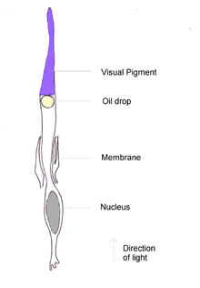
[10] Like rods, each cone cell has a synaptic terminal, inner and outer segments, as well as an interior nucleus and various mitochondria.

Photopigments exist as transmembrane proteins within these disks, which provide more surface area for light to affect the pigments.

Neither rods nor cones divide, but their membranous disks wear out and are worn off at the end of the outer segment, to be consumed and recycled by phagocytic cells.

The ratio of M and L cones varies greatly among different people with regular vision (e.g. values of 75.8% L with 20.0% M versus 50.6% L with 44.2% M in two male subjects).

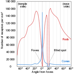
At lower light levels, where only the rod cells function, the sensitivity is greatest at a blueish-green wavelength.

The structure of a cone cell
Illustration of the distribution of cone cells in the fovea of an individual with normal color vision (left), and a color blind (protanopic) retina. Note that the center of the fovea holds very few blue-sensitive cones.
Distribution of rods and cones along a line passing through the fovea and the blind spot of a human eye [ 11 ]
1:posterior segment 2:ora serrata 3:ciliary muscle 4:ciliary zonules 5:Schlemm's canal 6:pupil 7:anterior chamber 8:cornea 9:iris 10:lens cortex 11:lens nucleus 12:ciliary process 13:conjunctiva 14:inferior oblique muscule 15:inferior rectus muscule 16:medial rectus muscle 17:retinal arteries and veins 18:optic disc 19:dura mater 20:central retinal artery 21:central retinal vein 22:optic nerve 23:vorticose vein 24:bulbar sheath 25:macula 26:fovea 27:sclera 28:choroid 29:superior rectus muscle 30:retina 1: posterior segment 2: ora serrata 3: ciliary muscle 4: ciliary zonules 5: Schlemm's canal 6: pupil 7: anterior chamber 8: cornea 9: iris 10: lens cortex 11: lens nucleus 12: ciliary process 13: conjunctiva 14: inferior oblique muscule 15: inferior rectus muscule 16: medial rectus muscle 17: retinal arteries and veins 18: optic disc 19: dura mater 20: central retinal artery 21: central retinal vein 22: optic nerve 23: vorticose vein 24: bulbar sheath 25: macula 26: fovea 27: sclera 28: choroid 29: superior rectus muscle 30: retina
1:posterior segment 2:ora serrata 3:ciliary muscle 4:ciliary zonules 5:Schlemm's canal 6:pupil 7:anterior chamber 8:cornea 9:iris 10:lens cortex 11:lens nucleus 12:ciliary process 13:conjunctiva 14:inferior oblique muscule 15:inferior rectus muscule 16:medial rectus muscle 17:retinal arteries and veins 18:optic disc 19:dura mater 20:central retinal artery 21:central retinal vein 22:optic nerve 23:vorticose vein 24:bulbar sheath 25:macula 26:fovea 27:sclera 28:choroid 29:superior rectus muscle 30:retina