Cyanosis

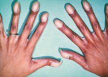
Cyanosis is the change of body tissue color to a bluish-purple hue, as a result of decrease in the amount of oxygen bound to the hemoglobin in the red blood cells of the capillary bed.

[1] Cyanosis is apparent usually in the body tissues covered with thin skin, including the mucous membranes, lips, nail beds, and ear lobes.

Furthermore, mongolian spots, large birthmarks, and the consumption of food products with blue or purple dyes can also result in the bluish skin tissue discoloration and may be mistaken for cyanosis.

While patients with increased amounts of red blood cells (e.g., polycythemia vera) can appear cyanotic even with lower concentrations of deoxyhemoglobin.

However, cyanosis can still be diagnosed with careful examination of the typical body areas such as nail beds, tongue, and mucous membranes where the skin is thinner and more vascular.

[5][6] Signs of severe anemia may include pale mucosa (lips, eyelids, and gums), fatigue, lightheadedness, and irregular heartbeats.

In patients with significant respiratory distress, supplemental oxygen (in the form of nasal canula or continuous positive airway pressure depending on severity) should be given immediately.

[12] It is postulated by Dr. Christen Lundsgaard that cyanosis was first described in 1749 by Jean-Baptiste de Sénac, a French physician who served King Louis XV.

[13] De Sénac concluded from an autopsy that cyanosis was caused by a heart defect that led to the mixture of arterial and venous blood circulation.

Presentation of cyanosis varies depending on the color of the skin
A baby with a heart condition. Note purple nailbeds.
Child with congenital heart disease with central cyanosis that is worsened by measles . Note the bluish-purple discoloration of the fingernails, lips, eyelids, and nose, along with prominent nail clubbing .
Peripheral cyanosis in an individual with peripheral vascular disease .
Initial direction of blood flow in patients with patent ductus arteriosus. Once the pressure of the pulmonary arteries increases more than the aorta due to right heart hypertrophy , the direction of blood flow reverses, sending deoxygenated blood through the patent duct directly into the descending aorta while sparing the brachiocephalic trunk , left common carotid , and left subclavian artery , therefore causing the differential cyanosis.
This illustration depicts a self-induced local (tissue) hypoxia on the right hand (right side of the picture) versus a normal left hand (left side of the picture). The cyanosis was achieved by inflating and tightening the blood pressure cuff on the right arm.
An example of cyanosis in an individual with darker skin pigmentation. Note the pale purple (instead of the typical bluish-purple hue) nail beds. This patient also had prominent digital clubbing due to a congenital heart disease with right-to-left shunting (this patient had Tetralogy of Fallot ).
An example of cyanosis in an elderly individual with darker skin pigmentation. Note the dark purple hue of the lips.