Cytogenetics
The next stage took place after the development of genetics in the early 20th century, when it was appreciated that the set of chromosomes (the karyotype) was the carrier of the genes.
[4] In 1912, Hans von Winiwarter reported 47 chromosomes in spermatogonia and 48 in oogonia, concluding an XX/XO sex determination mechanism.
Joe Hin Tjio working in Albert Levan's lab[8][9] was responsible for finding the approach: It took until 1956 for it to be generally accepted that the karyotype of man included only 46 chromosomes.
McClintock, while at the Carnegie Institution, continued previous studies on the mechanisms of chromosome breakage and fusion flare in maize.
[14] McClintock continued her career in cytogenetics studying the mechanics and inheritance of broken and ring (circular) chromosomes of maize.
During her cytogenetic work, McClintock discovered transposons, a find which eventually led to her Nobel Prize in 1983.
In the 1930s, Dobzhansky and his coworkers collected Drosophila pseudoobscura and D. persimilis from wild populations in California and neighboring states.
By the time Dobzhansky published the third edition of his book in 1951[16] he was persuaded that the chromosome morphs were being maintained in the population by the selective advantage of the heterozygotes, as with most polymorphisms.
Hotta, Chandley et al.[19] presented the evidence for a common pattern of DNA nicking and repair synthesis in male meiotic cells of lilies and rodents during the zygotene–pachytene stages of meiosis when crossing over was presumed to occur.
Abnormalities arising from nondisjunction events can cause cells with aneuploidy (additions or deletions of entire chromosomes) in one of the parents or in the fetus.
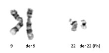
[citation needed] Acquired cytogenetics: In 1960, Peter Nowell and David Hungerford[21] discovered a small chromosome in the white blood cells of patients with Chronic myelogenous leukemia (CML).
The increasing knowledge of these cancer genes now allows the development of targeted therapies, which transforms the prospects of patient survival.
In the late 1960s, Torbjörn Caspersson developed a quinacrine fluorescent staining technique (Q-banding) which revealed unique banding patterns for each chromosome pair.
G-banding (utilizing trypsin and Giemsa/ Wright stain) was concurrently developed in the early 1970s and allows visualization of banding patterns using a bright field microscope.
These maps became the basis for both prenatal and oncological fields to quickly move cytogenetics into the clinical lab where karyotyping allowed scientists to look for chromosomal alterations.
[citation needed] High-resolution banding involves the staining of chromosomes during prophase or early metaphase (prometaphase), before they reach maximal condensation.
[23] Cells from bone marrow, blood, amniotic fluid, cord blood, tumor, and tissues (including skin, umbilical cord, chorionic villi, liver, and many other organs) can be cultured using standard cell culture techniques in order to increase their number.
After the cells have been allowed to sit in hypotonic solution, Carnoy's fixative (3:1 methanol to glacial acetic acid) is added.
The results are summarized and given to a board-certified cytogeneticist for review, and to write an interpretation taking into account the patient's previous history and other clinical findings.
The slides are then washed to remove the excess unbound probe, and counterstained with 4',6-Diamidino-2-phenylindole (DAPI) or propidium iodide.
[citation needed] Advances now focus on molecular cytogenetics including automated systems for counting the results of standard FISH preparations and techniques for virtual karyotyping, such as comparative genomic hybridization arrays, CGH and Single nucleotide polymorphism arrays.