Elbow
Deep fibres of the brachialis muscle insert anteriorly into the capsule and act to pull it and the underlying membrane during flexion in order to prevent them from being pinched.
A few of these fibres stretch across the olecranon fossa without attaching to it and form a transverse band with a free upper border.
They are positioned so that they always lie across the transverse joint axis and are, therefore, always relatively tense and impose strict limitations on abduction, adduction, and axial rotation at the elbow.
During rapid and forceful flexion all three muscles are brought into action assisted by the superficial forearm flexors originating at the medial side of the elbow.
Passive flexion (forearm is pushed against the upper arm with flexors relaxed) is limited to 160° by the bony projections on the radius and ulna as they reach to shallow depressions on the humerus; i.e. the head of radius being pressed against the radial fossa and the coronoid process being pressed against the coronoid fossa.
As the angle of flexion increases, the position of the olecranon approaches the main axis of the humerus which decreases muscle efficiency.
In full flexion, however, the triceps tendon is "rolled up" on the olecranon as on a pulley which compensates for the loss of efficiency.
Because triceps' long head is biarticular (acts on two joints), its efficiency is also dependent on the position of the shoulder.
Forced extension results in a rupture in one of the limiting structures: olecranon fracture, torn capsule and ligaments, and, though the muscles are normally left unaffected, a bruised brachial artery.
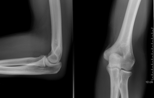
[14] The elbow undergoes dynamic development of ossification centers through infancy and adolescence, with the order of both the appearance and fusion of the apophyseal growth centers being crucial in assessment of the pediatric elbow on radiograph, in order to distinguish a traumatic fracture or apophyseal separation from normal development.
In humans, the main task of the elbow is to properly place the hand in space by shortening and lengthening the upper limb.
[21] Additionally, the forearm muscles that originate at the elbow are grouped at the sides of the joint in order not to interfere with its movement.
The wide angle of flexion at the elbow made possible by this arrangement — almost 180° — allows the bones to be brought almost in parallel to each other.
There is, however, extensive overlap in the carrying angle between individual men and women, and a sex-bias has not been consistently observed in scientific studies.
[26] Tennis elbow is the equivalent injury, but at the common extensor origin (the lateral epicondyle of the humerus).
A full dislocation of the elbow will require expert medical attention to re-align, and recovery can take approximately 6 weeks.
These repetitions can injure the tendons that connect the extensor supinator muscles (which rotate and extend the forearm) to the olecranon process (also known as "the elbow").
Wrist flexion and pronation (rotating of the forearm) causes irritation to the tendons near the medial epicondyle of the elbow.
[36] It can cause pain, stiffness, loss of sensation, and weakness radiating from the inside of the elbow to the fingers.
Ice, pain medication, steroid injections, strengthening exercises, and avoiding any aggravating activities can also help.
Exercises should focus on strengthening and stretching the forearm, and utilizing proper form when performing movements.
Most common treatments include wrist splints, surgery, physical and occupational therapy, and antirheumatic medication.
Other simple fixes include learning more ergonomically friendly habits that can help prevent nerve impingement and irritation in the future.
Recovery often includes movement restrictions, and range of motion activities, and can last a few months (cubital and radial tunnel syndrome, 2).
[44] Though the elbow is similarly adapted for stability through a wide range of pronation-supination and flexion-extension in all apes, there are some minor differences.
In arboreal apes such as orangutans, the large forearm muscles originating on the epicondyles of the humerus generate significant transverse forces on the elbow joint.
The structure to resist these forces is a pronounced keel on the trochlear notch on the ulna, which is more flattened in, for example, humans and gorillas.
In knuckle-walkers, on the other hand, the elbow has to deal with large vertical loads passing through extended forearms and the joint is therefore more expanded to provide larger articular surfaces perpendicular to those forces.
[45] Derived traits in catarrhini (apes and Old World monkeys), elbows include the loss of the entepicondylar foramen (a hole in the distal humerus), a non-translatory (rotation-only) humeroulnar joint, and a more robust ulna with a shortened trochlear notch.
In these taxa, the oval head of the radius lies in front of the ulnar shaft so that the former overlaps the latter by half its width.