Emergent virus
[13] Newly detected viruses may have escaped classification because they left no distinctive clues and/or could not be isolated or propagated in cell culture.
In order to overcome host-range restrictions and sustain efficient human-to-human transmission, viruses originating from an animal reservoir will normally undergo mutation, genetic recombination, and reassortment.
[20] Due to their rapid replication and high mutation rates, RNA viruses are more likely to successfully adapt for invasion of a new host population.
Their ability to fly and migration patterns also means that bats are able to spread disease over a large geographic area, while also acquiring new viruses.
[31] Factors such as deforestation, reforestation, habitat fragmentation, and irrigation can all impact the ways in which humans come into contact with wild animal species and consequently promote virus emergence.
Well-developed countries also have higher proportions of aging citizens and obesity-related disease, thus meaning that their populations may be more immunosuppressed and therefore at risk of infection.
[3] Contrastingly, poorer nations may have immunocompromised populations due to malnutrition or chronic infection; these countries are also unlikely to have stable vaccination program.
[3] Additionally, changes in human demographics[3] — for example, the birth and/or migration of immunologically naïve individuals — can lead to the development of a susceptible population that enables large-scale virus infection.
Other factors which can promote viral emergence include globalization; in particular, international trade and human travel/migration can result in the introduction of viruses into new areas.
[37][38] As hosts are immunologically naïve to pathogens they have not encountered before, emergent viruses are often extremely virulent in terms of their capacity to cause disease.
[citation needed] Although emergent viruses are frequently highly virulent, they are limited by several host factors including: innate immunity, natural antibodies, and receptor specificity.
[citation needed] Influenza is a highly contagious respiratory infection, which affects approximately 9% of the global population and causes 300,000 to 500,000 deaths annually.
[44][45] Influenza A viruses are further classified into subtypes, based on the combinations of the surface glycoproteins hemagglutinin (HA) and neuraminidase (NA).
The primary natural reservoir for most influenza A subtypes are wild aquatic birds;[44] however, through a series of mutations, a small subset of these viruses have adapted for infection of humans (and other animals).
Minor changes in HA and NA structure (antigenic drift) occur frequently, which enables the virus to cause repetitive outbreaks (i.e., seasonal influenza) by evading immune recognition.
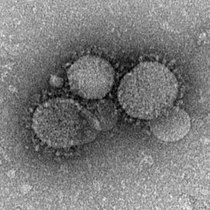
[44] In 2002, a highly pathogenic SARS-CoV (severe acute respiratory syndrome coronavirus) strain emerged from a zoonotic reservoir; approximately 8,000 people were infected worldwide, and mortality rates approached 50% or more in the elderly.
[51] The natural reservoir host for SARS-CoV-1 is thought to be horseshoe bats, although the virus has also been identified in several small carnivores (e.g., palm civets and racoon dogs).
The emergence of SARS-CoV-1 is believed to have been facilitated by Chinese wet markets, in which civets positive for the virus acted as intermediate hosts and passed SARS-CoV-1 onto humans (and other species).
[51] In order to infect cells, SARS-CoV-1 uses the spike surface glycoprotein to recognize and bind to host ACE-2, which it uses as a cellular entry receptor;[51] the development of this characteristic was crucial in enabling SARS-CoV-1 to 'jump' from bats to other species.