Endometrium
[1] The functional layer thickens and then is shed during menstruation in humans and some other mammals, including other apes, Old World monkeys, some species of bat, the elephant shrew[2] and the Cairo spiny mouse.
Vascular spaces fuse and become interconnected, forming the placenta, which supplies oxygen and nutrition to the embryo and fetus.
In the uterus, simple tubular glands reach from the endometrial surface through to the base of the stroma, which also carries a rich blood supply provided by the spiral arteries.
The well-documented presence of Lactobacillus species, for example, was easily explained by an increase in the vaginal population being able to seep into the cervical mucous.
However, in the uterus this much lower population is seen as invasive in a closed environment that is highly regulated by female sex hormones, and that could have unwanted consequences.
In studies of endometriosis Lactobacillus is not the dominant type and there are higher levels of Streptococcus and Staphylococcus species.
[7] The endometrium is the innermost lining layer of the uterus, and functions to prevent adhesions between the opposed walls of the myometrium, thereby maintaining the patency of the uterine cavity.
Vascular spaces fuse and become interconnected, forming the placenta, which supplies oxygen and nutrition to the embryo and fetus.
However, once ovulation occurs, the ovary (specifically the corpus luteum) will produce much larger amounts of progesterone.
Upon fertilization, the egg may implant into the uterine wall and provide feedback to the body with human chorionic gonadotropin (hCG).
If there is inadequate stimulation of the lining, due to lack of hormones, the endometrium remains thin and inactive.
The endometrium itself produces certain hormones at different stages of the cycle and this affects other parts of the reproductive system.
Chorionic tissue can result in marked endometrial changes, known as an Arias-Stella reaction, that have an appearance similar to cancer.
[18] The European Menopause and Andropause Society (EMAS) released Guidelines with detailed information to assess the endometrium.
Nevertheless, in human a perfect synchrony is not necessary; if the endometrium is not ready to receive the embryo an ectopic pregnancy may occur.
[24][25][26] Endometrial receptivity is a crucial factor in achieving successful embryo implantation in assisted reproduction treatments.
EMMA (Endometrial Microbiome Metagenomic Analysis) and ALICE (Analysis of Infectious Chronic Endometritis): Perform a test on the intrauterine microflora, using a small sample of the endometrium, in order to determine the presence of microorganisms that may promote or harm embryo implantation.

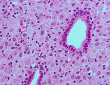

(A) proliferative endometrium (Left: HE × 400) and proliferative endometrial cells (Right: HE × 100)
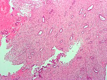
(B) secretory endometrium (Left: HE × 10) and secretory endometrial cells (Right: HE × 10)
(C) atrophic endometrium (Left: HE × 10) and atrophic endometrial cells (Right: HE × 10)
(D) mixed endometrium (Left: HE × 10) and mixed endometrial cells (Right: HE × 10)
(E): endometrial atypical hyperplasia (Left: HE × 10) and endometrial atypical cells (Right: HE × 200)
(F) endometrial carcinoma (Left: HE × 400) and endometrial cancer cells (Right: HE × 400).
