Lameness (equine)
[2] Metabolic causes of lameness include hyperkalemic periodic paralysis (HYPP) and polysaccharide storage myopathy, which directly affect muscular function.
Signs more commonly associated with a neurologic cause include unilateral muscle atrophy, paresis, paralysis, or dysmetria.
Generally, the viewer watches the hip, sacrum, gluteal muscles, or hemi-pelvis (pelvis of one side of the body) when examining lameness in the hind end.
If the pain is perceived during the early stance phase of the stride, the horse will try to unweight the leg and produce a "hip hike."
[17] Decreased height to the stride (flight arc), or dragging of the toes, also indicates lameness, as the horse avoids bending its joints.
It is the first step to evaluate decreased performance in an equine athlete, even if the horse does not appear overtly lame, to rule out any pain-associated cause.
Veterinarians may comment on aspects that could inhibit the use of the horse for the buyer's intended activity, such as subclinical osteoarthritis or conformational defects.
The pre-purchase exam may range in scope depending on the desire of the buyer, from a simple examination with hoof and flexion tests, to multiple radiographs, ultrasound, and advanced imaging techniques including MRI.
A good evaluation of conformation, including overall body type, can help the practitioner determine the potential cause of lameness.
Hind limb lameness or pelvic fracture can cause unilateral atrophy of the middle gluteal or gracilis muscles.
Neck muscle atrophy can be seen with cervical vertebral malformation (Wobbler's disease), articular facet osteoarthritis, and neurologic causes of lameness.
After a visual exam, the practitioner palpates the horse, feeling for heat, swelling, and sensitivity to pressure indicating pain.
[10] Specialized manipulative tests can be used to help identify specific areas of pain: The majority of lameness originates in the hoof.
For this reason, the hoof is closely scrutinized in shape, balance, shoeing, wear pattern, and for the presence of cracks, and contracted or sheared heels.
The process of watching a horse move is repeated after each additional flexion test or nerve block to determine its effect on the animal.
However, fractures and septic synovial structures (such as an infected joint pouch or tendon sheath) can also cause non-weight bearing lameness, and require emergency evaluation and treatment by a veterinarian.
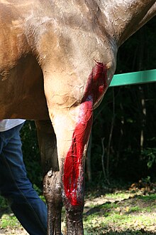
Therefore, non-weight bearing lameness should be assessed by an equine professional in a timely manner, especially if it is associated with trauma, laceration, or recent joint injection.
An increase in lameness following a flexion test suggests that those joints or surrounding soft tissue structures may be a source of pain for the horse.
Nerve blocks are performed in a step-wise fashion, beginning at the most distal (lower) part of the limb and moving upward.
False results can also be secondary to practitioner error if the anesthetic is accidentally administered into a location that was unintended, such as a synovial structure rather than around a nerve.
It is therefore a complementary imaging modality to radiographs, and is most commonly used to look for injury to ligaments and tendons, and the navicular bursa, although muscle damage and arterial blood flow have also been evaluated with ultrasound in cases of lameness.
The image may be manipulated to view in different planes, such as cross-section, making it possible to see an injury from multiple perspectives and improving diagnostic capabilities when compared to plain radiographs.
Advances in technology now also allows for a robotic scanner to rapidly image different parts the standing horse without the need for general anesthesia.
Like CT, an MRI image may be viewed in various planes of orientation, improving visualization of anatomic structures and any associated pathologic change.
However, standing MRI tends to be cheaper, and it eliminates the risks of general anesthesia, such as further damage to the injured area or additional injury that may occur during anesthetic recovery.
In adult horses, septic arthritis or tenosynovitis are most commonly seen secondary to joint injection, penetrating injury, or following surgery, and are often from Staphylococcus infection.
Arthroscopy is most commonly used for chip fractures of the knee and fetlock joints, osteochondritis dessecans lesions, and proliferative synovitis.
Appropriate treatment for lameness depends on the condition diagnosed, but at a minimum it usually includes rest or decreased activity and anti-inflammatory medications.
Other treatment options, such as corrective shoeing, joint injections, and regenerative therapies, are pursued based on the cause of lameness and the financial limits of the owner.
Consultation with a veterinarian is generally recommended, even for mild cases, as some types of lameness may worsen if not properly diagnosed and treated.