Eustachian tube
Active opening of the Eustachian tube (through actions like swallowing or the Valsalva maneuver) is required to equalize the pressure between the middle ear and the ambient atmosphere as the plane descends.
The mucous membrane of the tube is continuous in front with that of the nasal part of the pharynx, and behind with that of the tympanic cavity; it is covered with ciliated pseudostratified columnar epithelia and is thin in the osseous portion, while in the cartilaginous portion it contains many mucous glands and near the pharyngeal orifice a considerable amount of adenoid tissue, which has been named by Gerlach the tube tonsil.
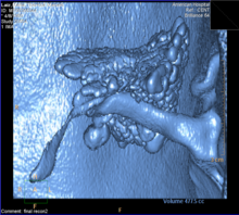
[9][10] Since 2015, two developments have enhanced our understanding of the anatomy of the eustachian tube: Valsalva computerized tomography and endoscopic ear surgery.
The distal part of the tubotympanic sulcus gives rise to the tympanic cavity, while the proximal tubular structure becomes the Eustachian tube.
Under normal circumstances, the human Eustachian tube is closed, but it can open to let a small amount of air through to prevent damage by equalizing pressure between the middle ear and the atmosphere.
Pressure differences cause temporary conductive hearing loss by decreased motion of the tympanic membrane and ossicles of the ear.
When this happens, humans hear a small popping sound, an event familiar to aircraft passengers, scuba divers, or drivers in mountainous regions.
In addition, children's developing immune systems and poor hygiene habits make them more prone to upper respiratory infections.
[citation needed] Barotitis, a form of barotrauma, may occur when there is a substantial difference in air or water pressure between the outer and the middle ear – for example, during a rapid ascent while scuba diving, or during sudden decompression of an aircraft at high altitude.
[17][unreliable medical source] It is suggested that Eustachian tube dysfunction can result in a large amount of mucus accumulating in the middle ear, often impairing hearing to a degree.
In severe cases of childhood middle ear infections and Eustachian tube blockage, ventilation can be provided by a surgical puncturing of the eardrum to permit air equalization, known as myringotomy.
[20][21][22] In the equids (horses) and some rodent-like species such as the desert hyrax, an evagination of the Eustachian tube is known as the guttural pouch and is divided into medial and lateral compartments by the stylohyoid bone of the hyoid apparatus.
This is of great importance in equine medicine as the pouches are prone to infections, and, due to their intimate relationship to the cranial nerves (VII, IX, X, XI) and the internal and external carotid artery, various syndromes may arise relating to which is damaged.