Tapetum lucidum

[8] The color in reindeer changes seasonally, allowing the animals to better avoid predators in low-light winter at the price of blurrier vision.

[3] Strepsirrhine primates are mostly nocturnal and, with the exception of several diurnal Eulemur species, have a tapetum lucidum of riboflavin crystals.

Its color is heterogeneous, varying with age and species due to factors like rodlet spacing, refractive index, and light interactions.

Young cats exhibit a blue appearance, which shifts to yellow with age, with adult coloration ranging from light orange to green.

[14] Kiwis, stone-curlews, the boat-billed heron, the flightless kākāpō, and many nightjars, owls, and other night birds such as the swallow-tailed gull possess a tapetum lucidum.

Four general patterns can be distinguished in spiders:[17] Animals without tapetum lucidum include haplorhine primates, squirrels, some birds, red kangaroo, and pigs.

In low light, a hand-held flashlight is sufficient to produce eyeshine that is visible to humans (despite their inferior night vision).

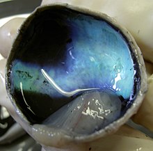
Eyeshine occurs in a wide variety of colors including white, blue, green, yellow, pink, and red.

However, since eyeshine is a type of iridescence, the color varies with the angle at which it is seen and the minerals which make up the reflective tapetum lucidum crystals.

Although human eyes lack a tapetum lucidum, they still exhibit a weak reflection from the choroid, as can be seen in photography with the red-eye effect and with near-infrared eyeshine.

[18][19] Another effect in humans and other animals that may resemble eyeshine is leukocoria, which is a white shine indicative of abnormalities such as cataracts and cancers.

[21] Traditionally, it has been difficult to take retinal images of animals with a tapetum lucidum because ophthalmoscopy devices designed for humans rely on a high level of on-axis illumination.

The dark blue, teal, and gold tapetum lucidum from the eye of a cow
Retina of a mongrel dog with strong tapetal reflex
Choroid dissected from a calf's eye, tapetum lucidum appearing iridescent blue
Reflection of camera flash from the tapetum lucidum
A three-month-old black Labrador puppy with apparent eyeshine
Reflective eyes of a cat visible from a camera flash
1:posterior segment 2:ora serrata 3:ciliary muscle 4:ciliary zonules 5:Schlemm's canal 6:pupil 7:anterior chamber 8:cornea 9:iris 10:lens cortex 11:lens nucleus 12:ciliary process 13:conjunctiva 14:inferior oblique muscule 15:inferior rectus muscule 16:medial rectus muscle 17:retinal arteries and veins 18:optic disc 19:dura mater 20:central retinal artery 21:central retinal vein 22:optic nerve 23:vorticose vein 24:bulbar sheath 25:macula 26:fovea 27:sclera 28:choroid 29:superior rectus muscle 30:retina 1: posterior segment 2: ora serrata 3: ciliary muscle 4: ciliary zonules 5: Schlemm's canal 6: pupil 7: anterior chamber 8: cornea 9: iris 10: lens cortex 11: lens nucleus 12: ciliary process 13: conjunctiva 14: inferior oblique muscule 15: inferior rectus muscule 16: medial rectus muscle 17: retinal arteries and veins 18: optic disc 19: dura mater 20: central retinal artery 21: central retinal vein 22: optic nerve 23: vorticose vein 24: bulbar sheath 25: macula 26: fovea 27: sclera 28: choroid 29: superior rectus muscle 30: retina
1:posterior segment 2:ora serrata 3:ciliary muscle 4:ciliary zonules 5:Schlemm's canal 6:pupil 7:anterior chamber 8:cornea 9:iris 10:lens cortex 11:lens nucleus 12:ciliary process 13:conjunctiva 14:inferior oblique muscule 15:inferior rectus muscule 16:medial rectus muscle 17:retinal arteries and veins 18:optic disc 19:dura mater 20:central retinal artery 21:central retinal vein 22:optic nerve 23:vorticose vein 24:bulbar sheath 25:macula 26:fovea 27:sclera 28:choroid 29:superior rectus muscle 30:retina