Lingual papillae
The four types of papillae on the human tongue have different structures and are accordingly classified as circumvallate (or vallate), fungiform, filiform, and foliate.
[2] They appear as very small, conical or cylindrical surface projections,[2] and are arranged in rows which lie parallel to the sulcus terminalis.
[2] Heavy keratinization of filiform papillae, occurring for instance in cats, gives the tongue a roughness that is characteristic of these animals.
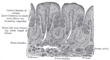
This epithelium has undergone a peculiar modification as the cells have become cone–like and elongated into dense, overlapping, brush-like threads.
[2] They are located on the sides at the back of the tongue, just in front of the palatoglossal arch of the fauces,[4][2] There are four or five vertical folds,[2] and their size and shape is variable.
Because their location is a high risk site for oral cancer, and their tendency to occasionally swell, they may be mistaken as tumors or inflammatory disease.
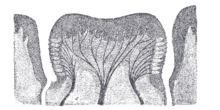
The papilla is shaped like a truncated cone, the smaller end being directed downward and attached to the tongue, the broader part or base projecting a little above the surface of the tongue and being studded with numerous small secondary papillae and covered by stratified squamous epithelium.
The function of the secretion is presumed to flush materials from the base of circular depression to ensure that taste buds can respond to changing stimuli rapidly.
In some diseases, there can be depapillation of the tongue, where the lingual papillae are lost, leaving a smooth, red and possibly sore area.
[4] Other sources state that foliate papilitis refers to inflammation of the lingual tonsil, which is lymphoid tissue.
Vallate (pronounced /ˈvæleɪt/ VAL-ayt) is from the Latin word vallum (rampart, wall), and means "having a raised edge surrounding a depression".
Foliate (pronounced /ˈfoʊliət/ FOH-lee-ət) is from the Latin word foliatus (leafy), and means "shaped like a leaf".