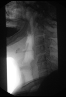
Fluoroscopy
In its primary application of medical imaging, a fluoroscope (/ˈflʊərəˌskoʊp/)[2][3] allows a surgeon to see the internal structure and function of a patient, so that the pumping action of the heart or the motion of swallowing, for example, can be watched.
The original difference was that radiography fixed still images on film, whereas fluoroscopy provided live moving pictures that were not stored.
Although visible light can be seen by the naked eye (and thus forms images that people can look at), it does not penetrate most objects (only translucent or transparent ones).
In contrast, X-rays can penetrate a wider variety of objects (such as the human body), but they are invisible to the naked eye.
(Depending upon what type of technology / panel is being used influences this answer greatly) Nowadays, in all forms of digital X-ray imaging (radiography, fluoroscopy, and CT) the conversion of X-ray energy into visible light can be achieved by the same types of electronic sensors, such as flat panel detectors, which convert the X-ray energy into electrical signals: small bursts of electric current that convey information that a computer can analyze, store, and output as images.
Flouroscopy will be use for each screw placed -which has greatly improved proper fracture heal due to more accurate reduction.
[5] In urology, fluoroscopy is used in retrograde pyelography and micturating cystourethrography to detect various abnormalities related to the urinary system.
In cardiology, fluoroscopy is used for diagnostic angiography, percutaneous coronary interventions, (pacemakers, implantable cardioverter defibrillators, and cardiac resynchronization devices).
Fluoroscopy's origins and radiography's origins can both be traced back to 8 November 1895, when Wilhelm Röntgen, or in English script Roentgen, noticed a barium platinocyanide screen fluorescing as a result of being exposed to what he would later call X-rays (algebraic x variable signifying "unknown").
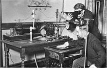
In the late 1890s, Thomas Edison began investigating materials for ability to fluoresce when X-rayed, and by the turn of the century he had invented a fluoroscope with sufficient image intensity to be commercialized.
Clarence Dally, a glass blower of lab equipment and tubes at Edison's laboratory was repeatedly exposed, developing radiation poisoning, later dying from an aggressive cancer.
[13] During this infant commercial development, many incorrectly predicted that the moving images of fluoroscopy would completely replace roentgenographs (radiographic still image films), but the then superior diagnostic quality of the roentgenograph and their already alluded-to safety enhancement of lower radiation dose via shorter exposure prevented this from occurring.
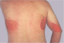
Issues raised by doctors and health professionals included the potential for burns to the skin, damage to bone, and abnormal development of the feet.
The development of the X-ray image intensifier by Westinghouse in the late 1940s[47] in combination with closed circuit TV cameras of the 1950s allowed for brighter pictures and better radiation protection.
Digital electronics were applied to fluoroscopy beginning in the early 1960s, when Frederick G. Weighart[48][49] and James F. McNulty[50] (1929–2014) at Automation Industries, Inc., then, in El Segundo, California produced on a fluoroscope the world's first image to be digitally generated in real-time, while developing a later commercialized portable apparatus for the onboard nondestructive testing of naval aircraft.
From the late 1980s onward, digital imaging technology was reintroduced to fluoroscopy after development of improved detector systems.
They include fluoroscopy, fluorography, cinefluorography, photofluorography, fluororadiography, kymography (electrokymography, roentgenkymography), cineradiography (cine), videofluorography, and videofluoroscopy.
Today, the word "fluoroscopy" is widely understood to be a hypernym of all the aforementioned terms, which explains why it is the most commonly used and why the others are declining in usage.
[51] The profusion of names is an idiomatic artifact of technological change, as follows: As soon as X-rays (and their application of seeing inside the body) were discovered in the 1890s, both looking and recording were pursued.
Another group of techniques included various kinds of kymography, whose common theme was capturing recordings in a series of moments, with a concept similar to movie film, although not necessarily with movie-type playback; rather, the sequential images would be compared frame by frame (a distinction comparable to tile mode versus cine mode in today's CT terminology).
Television also was under early development during these decades (1890s–1920s), but even after commercial TV began widespread adoption after World War II, it remained a live-only medium for a time.
Today, owing to technological convergence, the word "fluoroscopy" is widely understood to be a hypernym of all the earlier names for moving pictures taken with X-rays, both live and recorded.
Also owing to technological convergence, radiography, CT, and fluoroscopy are now all digital imaging modes using X-rays with image-analysis software and easy data storage and retrieval.
In the first half of the 20th century, shoe-fitting fluoroscopes were used in shoe stores, but their use was discontinued because it is no longer considered acceptable to use radiation exposure, however small the dose, for nonessential purposes.
Because fluoroscopy involves the use of X-rays, a form of ionizing radiation, fluoroscopic procedures pose a potential for increasing the patient's risk of radiation-induced cancer.
[63] Image intensifiers have been introduced that increase the brightness of the screen, so that the patient can be exposed to a lower dose of X-rays.
Flat-panel detectors offer increased sensitivity to X-rays, so have the potential to reduce patient radiation dose.
Spatial resolution is roughly equal, although an image intensifier operating in magnification mode may be slightly better than a flat panel.
A number of substances have been used as radiocontrast agents, including silver, bismuth, caesium, thorium, tin, zirconium, tantalum, tungsten, and lanthanide compounds.